Preparation method of metanephric mesenchyme cells

A cell and stem cell technology, applied in the fields of genetic engineering and molecular biology, can solve the problems of low cell yield, cumbersome steps, uncertain cell components, etc., and achieve the effect of single component, simple operation, and controllable extraction volume

Active Publication Date: 2016-12-21
GENERAL HOSPITAL OF PLA
View PDF3 Cites 2 Cited by
  • Summary
  • Abstract
  • Description
  • Claims
  • Application Information

AI Technical Summary

Problems solved by technology

[0003] The currently reported methods for isolating metanephric mesenchymal cells of a specific cell phenotype are not only cumbersome, but also have problems such as uncertain cell components and low cell yield.

Method used


Image

  • Preparation method of metanephric mesenchyme cells
  • Preparation method of metanephric mesenchyme cells
  • Preparation method of metanephric mesenchyme cells

Examples


Embodiment 1

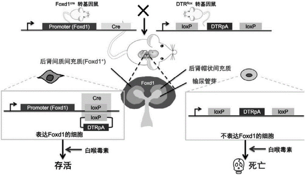
[0032] Example 1 Foxd1 + Preparation of metanephric mesenchymal cells

[0033] Foxd1 cre Transgenic mice with DTR flox The transgenic mice were mated, and whether the female mice were pregnant (whether there was a vaginal suppository) was observed twice a day in the morning and evening. If a vaginal suppository was found, it was recorded as E0.5 days, and the female mice were taken out and fed separately until the first E13.5-15.5 days. After the female rats were killed by neck dissection and disinfected, the uterus was immediately taken out, the embryos were separated, and the embryonic kidneys were separated with microsurgical instruments under a direct-view microscope. The embryonic kidneys of different embryonic mice were placed in EP tubes filled with normal saline, and The mouse tail corresponding to the embryonic mouse is also placed in the corresponding numbered EP tube to be used for genotype identification. Shred the embryonic kidneys and inoculate them in special...


PUM


Abstract

The invention provides a preparation method of metanephric mesenchyme cells. The method comprises the following steps: mating a Foxd1cre transgenic mouse with a DTRflox transgenic mouse, separating embryonic mouse kidneys, culturing embryonic kidney cells, and adding diphthera toxins to extract highly-pure Foxd1<+> metanephric mesenchyme cells. Compared with reported methods for separating specific cell phenotype metanephric mesenchyme cells, the method provided by the invention has the advantages of simplicity in operation, realization of simple components and high purity of the extracted Foxd1<+> metanephric mesenchyme cells, controllable extraction amount of the Foxd1<+> metanephric mesenchyme cells, and realization of large-scale production preparation of the Foxd1<+> metanephric mesenchyme cells.

Description

technical field [0001] The invention relates to the fields of genetic engineering and molecular biology, in particular to a preparation method of metanephric mesenchymal cells. Background technique [0002] The mammalian kidney develops from the interaction between metanephric ureteric buds and metanephric mesenchyme. The former develops into ureters and collecting ducts, and the latter can be divided into two groups of cells, namely cap mesenchymal cells (the marker is Six2 ) and interstitial mesenchymal cells (the marker is Foxd1). In the study of kidney development / embryonic kidney cells, it is sometimes necessary to isolate metanephric mesenchymal cells, or even metanephric mesenchymal cells of a specific cell phenotype. Cut, culture and isolate embryonic kidney cells, such as metanephric cells that require a specific cell phenotype (Foxd1 + Cells as an example), the isolated metanephric mesenchymal cells were stained with Foxd1 fluorescence, and then screened by flow ...

Claims


Application Information

Patent Timeline
IPC IPC(8): C12N5/073
CPCC12N5/0603C12N2509/00
Inventor 陈香美李清刚金美玲傅博
Owner GENERAL HOSPITAL OF PLA
Why Eureka
Try Eureka