A method for detecting the distribution of nanoparticles in cells and tissues and its application

A detection method and nanoparticle technology, applied in the field of medicine and biology, can solve the problems of fatigue, complex process, cumbersome and other problems, and achieve the effect of saving time, simple operation and more effective data.

Active Publication Date: 2020-02-28
SHANGHAI JIAOTONG UNIV
View PDF2 Cites 0 Cited by
  • Summary
  • Abstract
  • Description
  • Claims
  • Application Information

AI Technical Summary

Problems solved by technology

Conventional means for detection include flow cytometry and laser confocal microscopy, but the process is complex and cumbersome, and the data results provided cannot be quantified, and cannot accurately show the state of the nanoparticles themselves
Conventional instruments such as DLS for nanoparticle analysis also appear weak due to interference from serum proteins, blood cells, etc.

Method used


Image

  • A method for detecting the distribution of nanoparticles in cells and tissues and its application
  • A method for detecting the distribution of nanoparticles in cells and tissues and its application
  • A method for detecting the distribution of nanoparticles in cells and tissues and its application

Examples


Embodiment 1

[0062] Example 1 Quantitative detection of nanoparticle uptake by cells

[0063] (1) Preparation of fluorescent nanoparticles

[0064] The present invention uses liposomes as the model of nanoparticles. Take HSPC, Chol, and DSPE-PEG2000 stock solutions at a mass ratio of 3:1:1, add DiI at a lipid mass ratio of 1%, prepare liposomes by ethanol injection, hydrate with PBS for 30 minutes, and pass 200nm+ 100nm, 80nm+50nm Nuclepore polycarbonate membranes were passed 13 times each. To prevent fluorescence quenching, the preparation process was protected from light. The particle size of the prepared fluorescent liposomes was characterized by DLS.

[0065] (2) Cell uptake experiment

[0066] Plate after cell counting, incubate nanoparticles with a volume ratio of 1:1 with different media (PBS, albumin solution, pure serum), add cell culture medium, and incubate at 37°C for 2 hours, in which the concentration of albumin is 40mg / ml , the nanoparticle concentration was 1 mg / ml.

...

Embodiment 2

[0070] Example 2 Separation and Detection of Tissue Nanoparticles Methodological Investigation

[0071] (1) Preparation of fluorescent liposomes

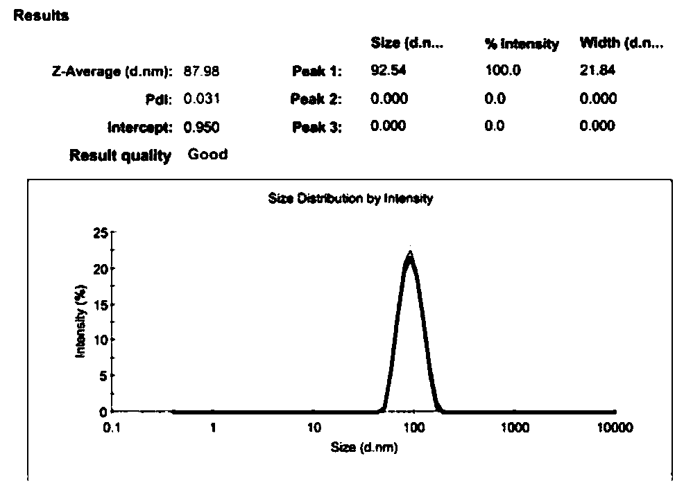
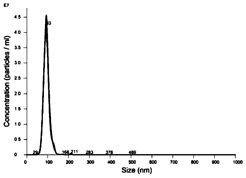
[0072] Take HSPC, Chol, and DSPE-PEG2000 stock solutions with a mass ratio of 3:1:1, add DiI at a lipid mass ratio of 1%, prepare liposomes by ethanol injection, hydrate with PBS for 30 minutes, and pass 200nm+ 100nm, 80nm+50nm Nuclepore polycarbonate membranes were passed 13 times each. In order to prevent fluorescence quenching, the preparation process was protected from light. Characterize the particle size of the prepared fluorescent liposomes with DLS, and the test results are as attached figure 2 shown.

[0073] (2) Isolation of nanoparticles in tissues

[0074] Cut 50mg of tumor tissue into pieces, add 50ul 6mg / ml, 0.6mg / ml, 0.06mg / ml fluorescent liposomes and mix well, add 750ul dispase solution (concentration is 2U / ml, U refers to a protease activity unit) , in a 37°C incubator, statically digest tumor tissue for 4 hou...

Embodiment 3

[0081] Example 3 Distribution detection of nanoparticles in tumors

[0082] (1) Animal experiments

[0083] The nude mice inoculated with tumors were randomly divided into 6 groups, one group was used as a blank control group, and the other 5 groups were injected with fluorescently labeled liposomes in the tail vein, and the mice were killed 2h, 6h, 9h, 12h, and 24h after administration, respectively. , the tumor tissue was obtained after dissection.

[0084] (2) Isolation of nanoparticles from tumor tissue

[0085] Weigh 50mg of tumor tissue at 2h, 6h, 9h, 12h, and 24h, add 750ul dispase solution (concentration is 0.6U / ml, U refers to a protease activity unit), and digest tumor tissue at 37°C for 4h , and then placed in a centrifuge, separated fluorescent liposomes by gradient centrifugation, centrifuged at 300g, 10min to remove intact cells; took the supernatant and centrifuged at 2000g, 10min to remove dead cells, cell debris, platelets, etc.; took the supernatant, passed...


PUM


Abstract

The invention belongs to the field of medicinal biology and particularly relates to a method of detecting distribution of nanoparticles in cells and tissues. A simple and reliable qualitative and / or quantitative detection method is provided by using nanoparticle tracking analysis technology to evaluate nanoparticle intake by cells as well as distribution of the nanoparticles in bodily tissues. By subjecting the nanoparticles to fluorescence labeling, the quantity and particle size of nanoparticles entering tissue organs are specifically analyzed by means of separation so as to investigate dynamic changes of the nanoparticles in a human body. The method of the invention has the advantages that operating is simple, time is saved, and much effective data are provided.

Description

technical field [0001] The invention belongs to the field of medicine and biology, in particular to a method for detecting the distribution of nanoparticles in cells and tissues and its application. Background technique [0002] After nanoparticles enter the body, complex interactions occur with proteins (albumin, lipoprotein, immunoglobulin, etc.) qualitative and quantitative changes. Conventional means for detection include flow cytometry and laser confocal microscopy, but the process is complex and cumbersome, and the data results provided cannot be quantified, and cannot accurately show the state of the nanoparticles themselves. Conventional instruments such as DLS for nanoparticle analysis also appear weak due to interference from serum proteins, blood cells, etc. The invention uses the nano particle tracking analysis technology to measure the particle diameter and particle number of the nano particle. At the same time, by fluorescently labeling specific nanoparticle...

Claims


Application Information

Patent Timeline
Patent Type & Authority Patents(China)
IPC IPC(8): G01N21/64
CPCG01N21/6486
Inventor 徐宇虹许风伟
Owner SHANGHAI JIAOTONG UNIV
Why Eureka
Try Eureka