A left ventricular volume reduction mechanism

A technology for the left ventricle and apex of the heart, applied in the fields of heart valves, medical science, surgery, etc., can solve the problems of inconvenient system introduction, affecting the quality of life of patients, increasing blood vessel and heart trauma, etc., to reduce the risk of fatigue fracture of the mechanism, and achieve a good three-dimensional Spatial vision and the effect of improving mechanical stability

Active Publication Date: 2018-11-02
NINGBO DIOCHANGE MEDICAL TECH CO LTD
View PDF8 Cites 0 Cited by
  • Summary
  • Abstract
  • Description
  • Claims
  • Application Information

AI Technical Summary

Problems solved by technology

The defects of this mechanism are: 1. According to the theory of heart torsion motion, in addition to the periodic systolic and diastolic motion in the radial direction (perpendicular to the central axis of the base), the heart will also undergo a circular rotation direction (along the direction of rotation) during contraction and relaxation. The periodic reciprocating torsional movement of the central axis of the base), after the base of the mechanism is released on the apex, it can only resist the periodic systolic and diastolic motions in the radial direction for buffering in the radial direction, but because the petal edges of the base are fixed by The connection is in a closed state and cannot resist the periodic reciprocating torsional movement in the direction of circular rotation, so it cannot produce a buffering effect in the direction of circular rotation, which in turn causes a series of problems, including the displacement of the mechanism due to being driven, the fatigue fracture of the base, the edge of the metal skeleton Anchor breakage has been reported in the clinical follow-up of this product
2. Since the base is petal-shaped as a whole, and the edges of the petals are in a closed state through fixed connections, the overall space occupied when it is installed into the delivery sheath is relatively large, so the delivery sheath is required to have a larger diameter, which increases the mechanism’s time of introduction. 3. After the mechanism is completely released, the blood in the heart will generate periodicity in the axial direction (parallel to the central axis of the base) The impact force impacts the center of the metal skeleton of the mechanism and then transmits it to the center of the base, and the flat base is difficult to disperse the impact force from the center of the base to the entire base, including the edge of the petals, so the base is in the axial direction and The effect of shock absorption is not really produced, which eventually leads to the fatigue fracture of the metal skeleton in the central area where the metal skeleton gathers. At present, relevant reports have been reported in the clinical follow-up of this product
The base of the mechanism still has the same problems as the base of the Parachute left ventricle volume reduction mechanism, namely: 1. According to the theory of heart torsion motion, the heart is in the process of systole and diastole except for the cycle in the radial direction (perpendicular to the central axis of the base) systolic and diastolic movements, and periodic reciprocating torsional movements in the direction of circular rotation (rotating along the central axis of the base). Cushioning in the radial direction, but because the edge of the petals of the base is in a closed state through a fixed connection, it cannot resist the periodic reciprocating torsional movement in the direction of circular rotation, so the buffering effect in the direction of circular rotation cannot be produced, which in turn causes a series of problems. Driven and shifted, the fatigue of the base breaks, and the anchor thorns on the edge of the metal skeleton break; 2. Since the blood in the heart generates periodic impact force in the axial direction (parallel to the central axis of the base), the main body of the impact mechanism is collected by this force. The center is then transmitted to the base. The base is designed with a cross-woven structure of metal wires and other materials. Although it can play a shock-absorbing role in the axial direction, the metal wires will continue to rub against each other under the action of periodic impact forces, which is extremely It is easy to cause fatigue fracture; 3. Furthermore, the woven cage structure base causes the overall length of the mechanism to be too long, which increases the rigidity of the system, which is not convenient for the system to be introduced in the tortuous blood vessel and cardiac access system, and also brings inconvenience to the operation. It is extremely difficult to operate and requires a large space during the intervention, which may increase the risk of trauma to blood vessels and the heart
[0011] To sum up, the existing left ventricular volume reduction mechanism cannot completely solve the problem of fatigue fracture and displacement of the mechanism caused by the torsional movement of the heart
Seriously affect the quality of life of patients after surgery, and even affect the life expectancy, which shows that the safety and effectiveness of institutions need to be improved urgently

Method used


Image

  • A left ventricular volume reduction mechanism
  • A left ventricular volume reduction mechanism
  • A left ventricular volume reduction mechanism

Examples


Embodiment 1

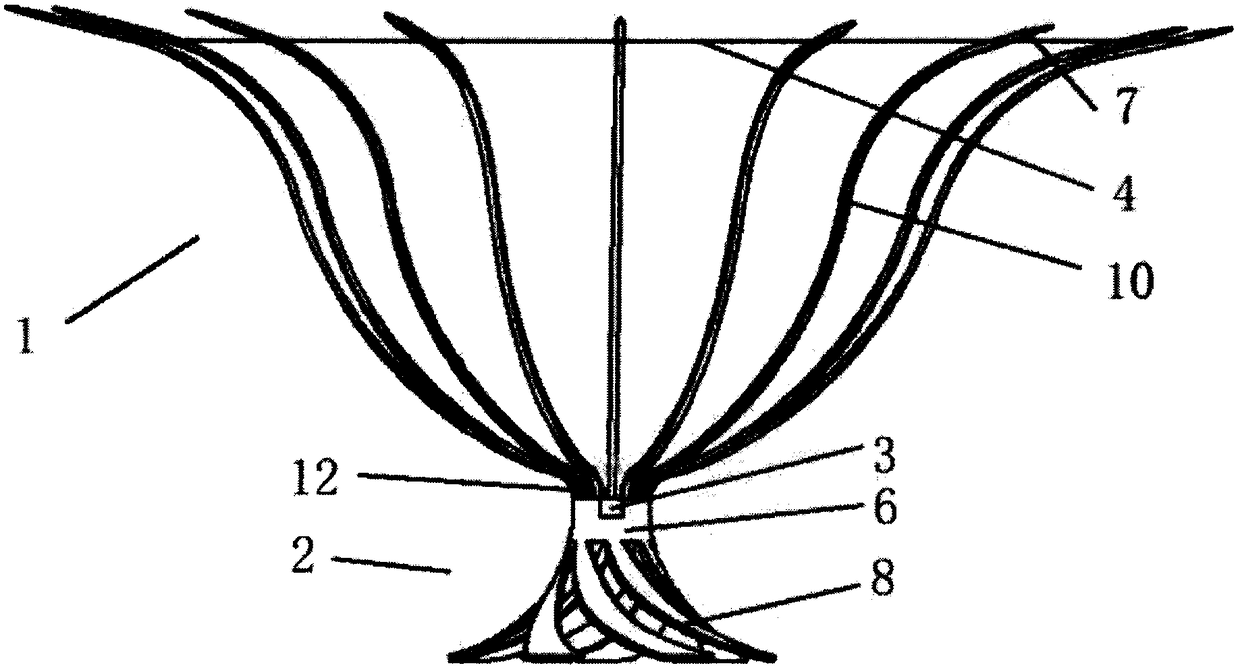
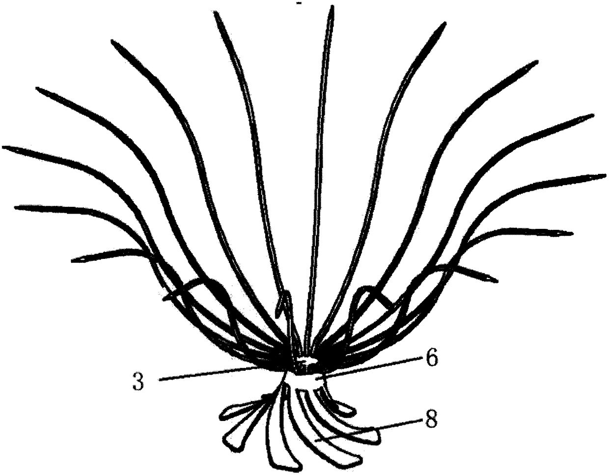
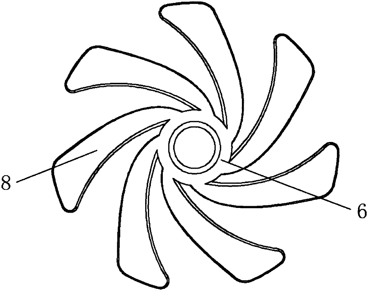
[0055] Such as Figures 1 to 4 As shown, a left ventricular volume reduction mechanism includes a support frame 1 and a base 2 connected to the support frame 1; the support frame 1 includes a plurality of support rods 10 that are adaptively expanded away from the apex 5, and the plurality of support rods 10 are The proximal ends of the support rods 10 are merged together to form a gathered bottom end 12, and the distal ends of the support rods 10 are provided with anchors 7. When the support frame 1 is released in the left ventricle 9, the support rods 10 are adaptively close to the inner wall of the ventricle , the anchor thorn 7 pierces the inner wall of the ventricle to anchor the left ventricular volume reduction mechanism; the base 2 includes a central end 6 connected to the gathered bottom end 12, and extends from the central end 6 toward the apex A plurality of helical branches 8, the plurality of helical branches 8 are rotationally symmetrical along the central axis of...

Embodiment 2

[0064] Such as Figure 5 and Figure 6 As shown, on the basis of embodiment 1, the difference between embodiment 2 and embodiment 1 is that the end of the helical branch 8 near the apex 5 is provided with a crimp 81, in one embodiment, the crimp 81 is that the proximal end of the helical branch 8 extends smoothly and then curls into a closed circle or hook shape toward the axial direction of the base 2 . The crimp 81 abuts against the inner wall of the apex 5 . This design has many advantages, including: it is convenient for the mechanism to be easily pushed out from the delivery sheath tube to prevent hanging the inner wall of the sheath; the visualization during the operation is enhanced, and it is convenient for positioning judgment when the mechanism is released; after the mechanism is released in the left ventricle 9, both It can be smoothly adapted to the inner wall of the apex 5, and can also reduce the torsion force generated by the twisting movement of the heart on ...

Embodiment 3

[0067] Such as Figure 7 As shown, on the basis of Embodiment 1, the difference between Embodiment 3 and Embodiment 1 is that the proximal end of the helical branch 8 is provided with a ball head 82. This design has many advantages, including: Easily push out from the delivery sheath to prevent the inner wall of the sheath from hanging; enhance the visualization during the operation, and facilitate the positioning judgment when the mechanism is released; after the mechanism is released in the left ventricle 9, it can be smoothly adapted to the inner wall of the apex 5, or Reduce the torsion force generated by the heart torsion movement on the mechanism, prevent the mechanism from shifting, and reduce side leakage and other complications. In addition, a through hole 88 can be set in the ball head 82, such as Figure 8 As shown in the partial cross-sectional view A of , the through hole 88 is used to pass through the series member 86 .


PUM


Abstract

The invention relates to a left ventricular volume reduction mechanism, which includes a support frame and a base connected to the support frame. The support frame is composed of a plurality of support rods, and the proximal ends of the support rods are gathered to form a gathering of the support frame. The bottom end has a distal end that is adaptively opened along the inner wall of the left ventricle in a direction away from the apex. The base includes a central end connected to the gathered bottom end, and a plurality of spirals extending from the central end toward the apex. Branches, a plurality of the spiral branches are rotationally symmetrical along the central axis of the base, and the entire body forms a trumpet shape with the opening facing the apex direction. The support frame is made of shape memory material and its surface is covered with a film. The base is made of Made of elastic material, the base adaptively fits the inner wall of the apex. The invention reduces the risk of instrument displacement and breakage, has good fatigue strength, and reduces the risk of damaging blood vessels and heart valves.

Description

technical field [0001] The invention relates to the technical field of medical ventricular isolation devices, in particular to a left ventricular volume reduction mechanism. Background technique [0002] In recent years, the prevalence of heart failure has been increasing year by year, becoming a serious public health problem. Patients with clinical symptoms are similar to malignant tumors. [0003] It has many causes, one of the most common causes is acute myocardial infarction. Clinically, especially in patients with anterior wall myocardial infarction and ventricular aneurysm, the curvature and thickness of the left ventricular wall can be altered, resulting in dilation and deformation of the entire heart. Since the aneurysm affects systolic function, paradoxical motion of the aneurysm portion reduces left ventricular ejection. Combined with scar formation after myocardial infarction, changes in the intrinsic properties of the myocardium and marginal tissue increase the...

Claims


Application Information

Patent Timeline
Patent Type & Authority Patents(China)
IPC IPC(8): A61B17/00A61B17/12A61F2/24
CPCA61B17/0057A61B17/12022A61B17/12122A61B17/1215A61F2/2487A61B2017/12095A61B2017/00243A61B2017/00615A61B2017/00592A61B2017/00575A61B2017/00632A61B2017/00867A61B17/1214
Inventor 李欣欣葛均波李彪吕世文周达新沈雳
Owner NINGBO DIOCHANGE MEDICAL TECH CO LTD
Why Eureka
Try Eureka