Inflammatory myopathy SAE1 self antibody non-radioactive detection method and application thereof

An autoantibody and detection method technology, applied in the biological field, can solve the problems of high cost and high false positive rate of ELISA kits

Inactive Publication Date: 2017-04-26
CENT SOUTH UNIV
View PDF3 Cites 1 Cited by
  • Summary
  • Abstract
  • Description
  • Claims
  • Application Information

AI Technical Summary

Problems solved by technology

[0007] In order to overcome the defects of the above-mentioned existing technologies, and solve the defects of ELISA kits and western blot membrane strips that are not commercially available in China for detecting SAE1 antibodies, as well as the high cost, high false positive rate and radiolabeling of ELISA kits that self-coat SAE1 antigens Reg

Method used


Image

  • Inflammatory myopathy SAE1 self antibody non-radioactive detection method and application thereof
  • Inflammatory myopathy SAE1 self antibody non-radioactive detection method and application thereof
  • Inflammatory myopathy SAE1 self antibody non-radioactive detection method and application thereof

Examples


Embodiment 1

[0028] Example 1 Non-radiolabeled immunoprecipitation detection method for SAE1 autoantibodies in inflammatory myopathy

[0029] 1. Materials

[0030] pENTER-SAE1 plasmid (Virgin Bio, CH838665)

[0031] Flag antibody (Sigma-Aldrich, F1804)

[0032] Protein G Protein G magnetic beads (Millipore, LSKMAGG10)

[0033] 2. Method

[0034] A non-radiolabeled immunoprecipitation detection method for SAE1 autoantibodies in inflammatory myopathy, comprising the following steps:

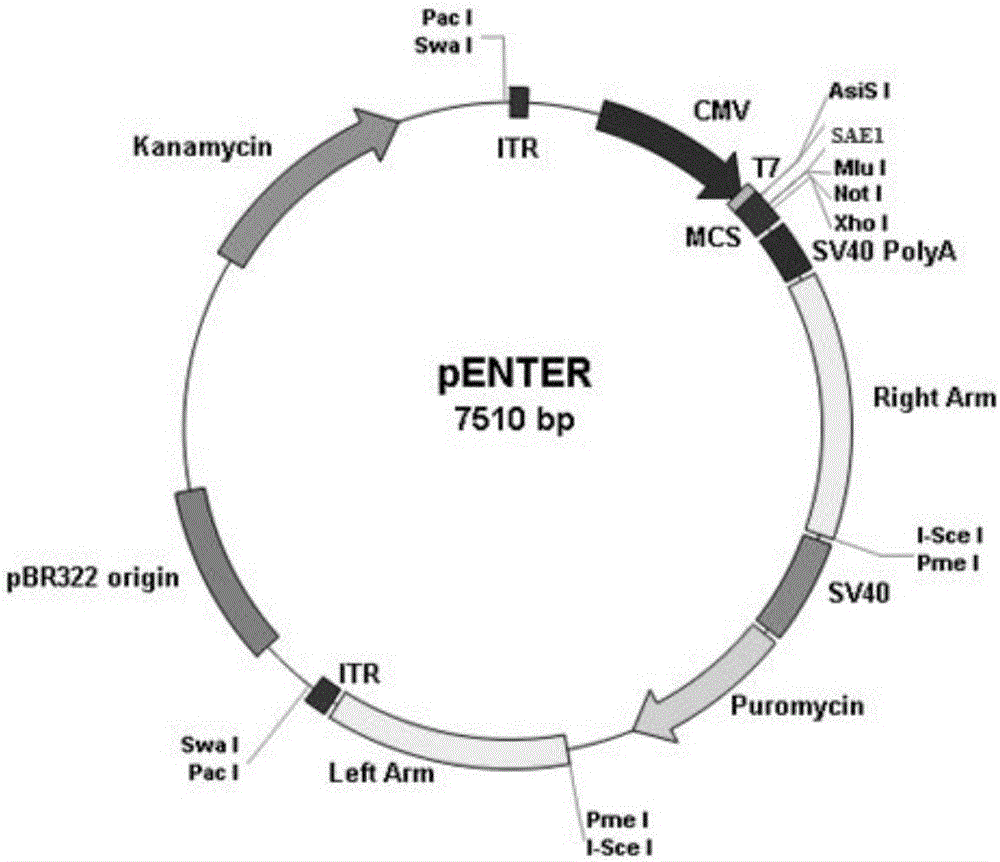
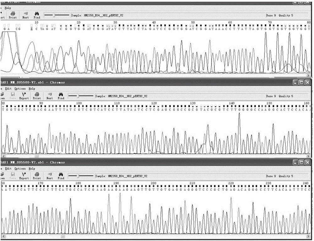
[0035] (1) The pENTER-SAE1 plasmid with the Flag tag (as attached figure 1 shown) for sequencing to confirm the accuracy of the SAE1 sequence and reading frame (as attached figure 2 shown);

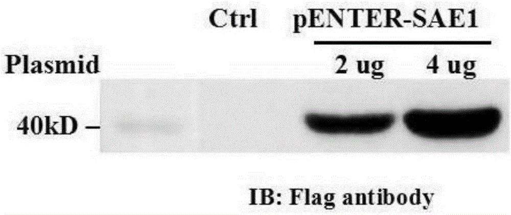
[0036] (2) 2ug pENTER-SAE1 and 4ug pENTER-SAE1 were transiently transfected into human embryonic kidney (HEK) 293 cells for 48 hours, and the HEK293 cells were lysed with SDS lysate, and then SDS-PAGE gel electrophoresis and membrane transfer were performed using Flag antibody. After incubation, it was found that t...

Embodiment 2

[0038] Example 2 Western blot detection method for inflammatory myopathy SAE1 autoantibody

[0039] 1. Materials

[0040] pENTER-SAE1 plasmid (Virgin Bio, CH838665)

[0041] Flag antibody (Sigma-Aldrich, F1804)

[0042] Protein G Protein G magnetic beads (Millipore, LSKMAGG10)

[0043] SAE1 antibody positive serum (1:100 dilution)

[0044] Rabbit anti-human IgG H&L (1:5000, Abcam, ab6759))

[0045] 2. Method

[0046] The immunoblotting method for detecting SAE1 autoantibodies in inflammatory myopathy comprises the following steps:

[0047] (1) 2ug pENTER-SAE1 and 4ug pENTER-SAE1 were transiently transfected into HEK293 cells for 48 hours respectively, and the HEK293 cells were lysed with SDS lysate, then subjected to SDS-PAGE gel electrophoresis and membrane transfer, and incubated with Flag antibody, and it was found that transfected pENTER - There is a specific band of about 40kDa in the HEK293 cells of the SAE1 plasmid (as attached image 3 shown), the above results ...

Embodiment 3

[0049] Example 3 detects the positive rate of SAE1 antibody in the serum of 52 patients with inflammatory myopathy

[0050] The positive rate of SAE1 antibody in the serum of patients with inflammatory myopathy includes the following steps:

[0051] (1) Inoculate 30% HEK293 cells on 75cm 2 Cell culture dish, cultivated at 37°C for less than 24 hours;

[0052] (2) Transfect HEK293 cells with 20ug pENTER-SAE1 plasmid for 48 hours;

[0053] (3) HEK293 cells were lysed with 1 ml of RIPA lysate containing protease inhibitors, and the protein was quantified;

[0054] (4) Using Flag antibody to confirm the overexpression of SAE1 in HEK293 cells;

[0055] (5) Take 20ul of the test serum, positive serum, and negative serum from patients with inflammatory myopathy to perform immunoprecipitation with the cell lysate overexpressing SAE1, perform SDS-PAGE gel electrophoresis, and then detect with Flag antibody to determine the SAE1 antibody The positive rate in patients with inflammato...


PUM


Abstract

The invention discloses an inflammatory myopathy SAE1 self antibody non-radioactive detection method and an application thereof. The method comprises the following steps: an inflammatory myopathy SAE1 self antibody non-radioactive mark immunoprecipitation detection method and an immunoblotting detection method which are used for detecting an anti-SAE1 self antibody of the patients with idiopathic inflammatory myopathy; the non-radioactive mark immunoprecipitation detection method comprises the following steps: using Protein G magnetic bead for respectively combining with the SAE1 antibody positive inflammatory myopathy serum or health contrast serum, then performing incubation with over-expressed SAE1 HEK293 cell lysis buffer for antigen adsorption, after elution, performing SDS-PAG gel electrophoresis, and using a Flag antibody for detection; the immunoblotting detection method comprises the following steps: performing SDS-PAG gel electrophoresis on the over-expressed SAE1 HEK293 cell lysis buffer, using SAE1 antibody positive serum a primary antibody, and using rabbit anti human IgG H&l as a secondary antibody for detection. According to the invention, problems of an ELISA kit for detecting SAE1 antibody and immunization immunization blotting membrane without commercialization in our country can be solved, and the problems of high cost of the ELISA kit for self-coating of the SAE1 antigen, high false positive rate and security of a radioactive mark immunoprecipitation method can be overcome.

Description

technical field [0001] The application belongs to the field of biotechnology, and relates to a non-radioactive detection method and application of SAE1 autoantibodies in inflammatory myopathy, in particular to a non-radioactive labeling immunoprecipitation detection method and immunoblotting detection method and application of SAE1 autoantibodies in inflammatory myopathy. Background technique [0002] Idiopathic inflammatory myopathy (idiopathic inflammatory myopathies, IIM) is a group of systemic autoimmune diseases mainly invading skeletal muscle, the abnormality of autoimmunity is the key to the occurrence and development of IIM. According to the latest diagnostic classification criteria, IIM can be divided into polymyositis (PM), dermatomyositis (DM), immune-mediated necrotizing myopathy (IMNM), nonspecific Myositis (nonspecific myositis, NSM) and inclusion body myositis (inclusion body myositis, sIBM). The clinical features of IIM are muscle weakness in the proximal ex...

Claims


Application Information

Patent Timeline
IPC IPC(8): G01N33/68G01N33/564
CPCG01N33/564G01N33/6854G01N2800/10G01N2800/24
Inventor 张华莉王莉黄莉罗卉李懿莎左晓霞朱红林王国春刘瑛贺维佳刘可刘梅冬陈广文肖献忠
Owner CENT SOUTH UNIV
Why Eureka
Try Eureka