Colorectal cancer liver metastasis animal model construction method for multi-modal imaging research

A multi-modal imaging and colorectal cancer technology, applied in the fields of application, medical science, surgery, etc., can solve the problems of long induction period, hepatic vascular embolism, and short survival time of experimental animals, and achieve simple operation and high modeling success rate high effect

Inactive Publication Date: 2017-05-10
HARBIN MEDICAL UNIVERSITY
View PDF0 Cites 1 Cited by
  • Summary
  • Abstract
  • Description
  • Claims
  • Application Information

AI Technical Summary

Problems solved by technology

The disadvantages of this model are: (1) the whole process of tumor metastasis could not be simulated, and the initial steps of primary tumor invasion into surrounding tissue and penetration into blood vessels were reduced; (2) the survival time of experimental animals was relatively short; (3)

Method used


Image

  • Colorectal cancer liver metastasis animal model construction method for multi-modal imaging research
  • Colorectal cancer liver metastasis animal model construction method for multi-modal imaging research
  • Colorectal cancer liver metastasis animal model construction method for multi-modal imaging research

Examples


Embodiment 1

[0040] Experimental animals and materials, equipment

[0041] Experimental animal: the animal model of liver metastasis of colorectal cancer constructed in Example 1.

[0042] Anhydrous ether: Tianjin Fuyu Fine Chemical Co., Ltd.

[0043] Ultrasound equipment: Model: PHILIPS HD15, produced by Philips Company in the Netherlands.

[0044] experimental method

[0045] Carry out liver ultrasound scanning on the colorectal cancer liver metastasis nude mouse model constructed in the present invention, the operation is as follows:

[0046] (1) The examiner wears sterile gloves, anesthetizes the nude mice in the experimental group with anhydrous ether, and fixes the nude mice on the self-made inspection board with medical tape after anesthesia.

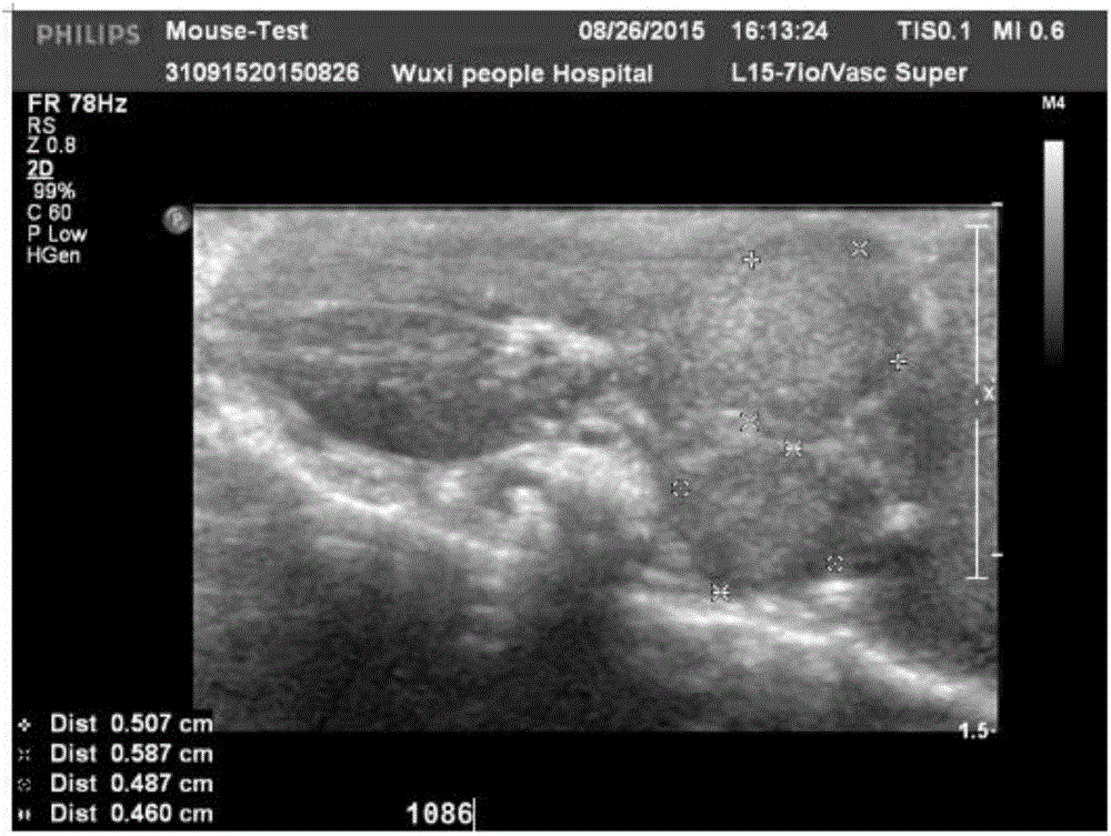
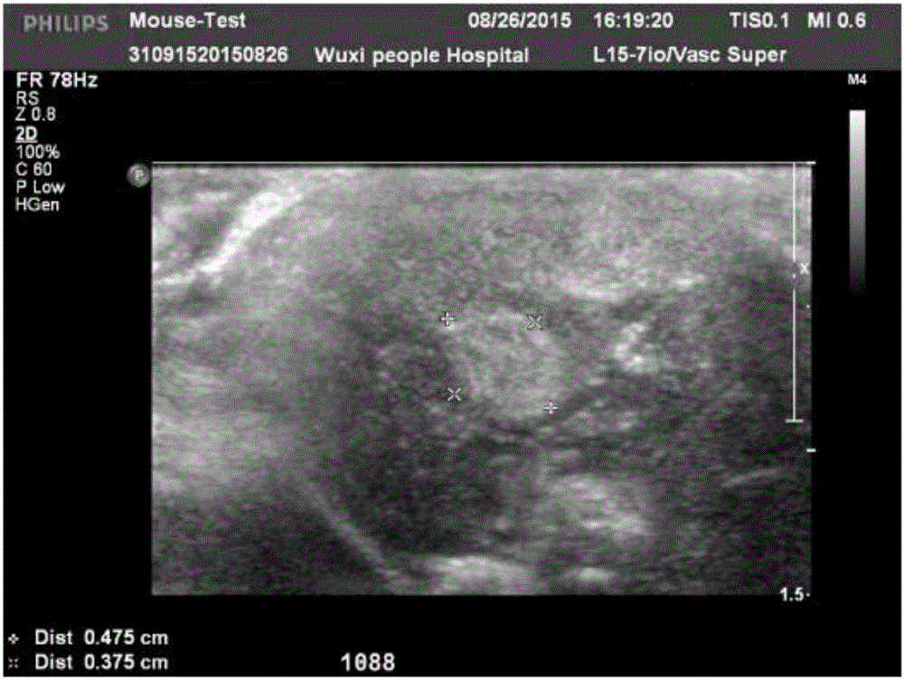
[0047] Ultrasound equipment was used to scan the nude mice. The probe used was the L15-7io superficial probe. The abnormal echoes of the liver were recorded, and the images were collected after measuring the size.

[0048] Ultrasound exam...

Embodiment 2

[0050] Experimental animals and materials, equipment

[0051] Experimental animal: the animal model of liver metastasis of colorectal cancer constructed in Example 1.

[0052] Isoflurane: Shanghai Abbott Pharmaceutical Co., Ltd.; Oxygen: Wuxi Liyuan Medical Oxygen Co., Ltd. Small animal anesthesia machine: Stoelting Kopf / 50262, USA; Micro-CT: Siemens, Germany.

[0053] experimental method

[0054] Liver scanning was performed on the colorectal cancer liver metastasis nude mouse model constructed in the present invention, and the operation was as follows:

[0055] (1) Before imaging, nude mice were anesthetized with oxygen mixed with 3% (volume fraction) isoflurane on a small animal anesthesia machine, and maintained oxygen anesthesia containing 1% (volume fraction) isoflurane during the imaging process, in prone position The advanced position of the head is fixed on the scanning table.

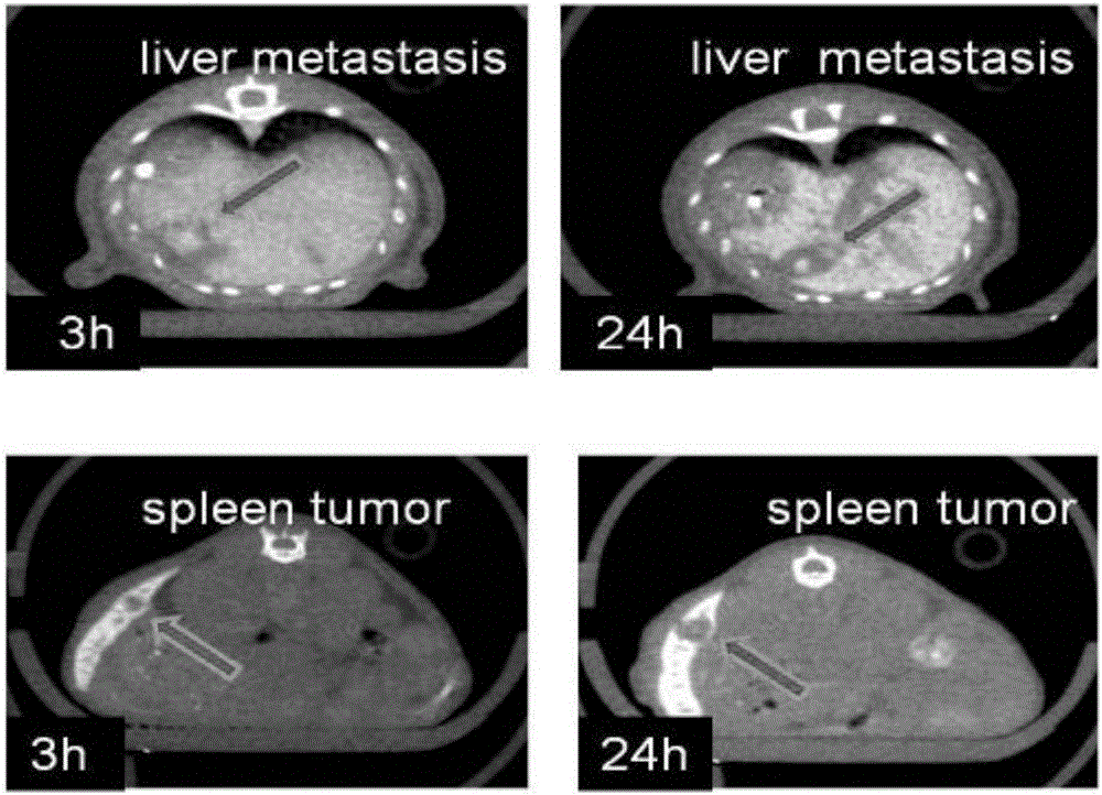
[0056] (2) The contrast agent used was Fenestra LC, and the amount of contrast agent w...

Embodiment 3

[0060] Experimental animals and materials, equipment

[0061] Experimental animal: the animal model of liver metastasis of colorectal cancer constructed in Example 1.

[0062] Isoflurane: Shanghai Abbott Pharmaceutical Co., Ltd.; Oxygen: Wuxi Liyuan Medical Oxygen Co., Ltd.

[0063] Small animal anesthesia machine: American Stoelting Kopf / 50262; Micro-MRI: model BRUKER 7.0TBiospec 70 / 20USR.

[0064] experimental method

[0065] The liver metastases nude mouse model of colorectal cancer constructed in Example 1 was subjected to magnetic resonance liver transverse and coronal scans, and the operation was as follows:

[0066] (1) Before imaging, nude mice were anesthetized with oxygen mixed with 3% (volume fraction) isoflurane on a small animal anesthesia machine, and maintained oxygen anesthesia containing 1% (volume fraction) isoflurane during the imaging process, in prone position The advanced position of the head is fixed on the scanning table.

[0067] (2) Nuclear magnet...


PUM


Abstract

The invention discloses a colorectal cancer liver metastasis animal model construction method for multi-modal imaging research. The defect that an existing colorectal cancer liver metastasis implanting model is high in colorectal obstruction morbidity and short in model survival time and cannot adapt to long-term experiment is overcome. Adopted animals are 5-year-old BALB/C-nu-nu female nude mice, an adopted injection is human colorectal cancer cells (HT29), and the method comprises the construction steps that 0.1 ml of colorectal cancer cells with the concentration being 2.5*10<7>/ml prepared from 0.01 MPBS is injected into the spleen, the injection time is about 3 min, the spleen is placed into an enterocoelia after injection is finished, and a colorectal cancer liver metastasis nude mouse model can be obtained seven weeks after the model is manufactured. Due to the fact that the concentration of the colorectal cancer cells (HT29) is 25*10<7>/ml, the dosage of the colorectal cancer cells is 0.1 ml, the liver metastasis model can be obtained by only injecting a cell suspension into the spleen at a time, and therefore the method is easy to implement, high in molding success rate and worthy of popularization and has certain economic and practical value.

Description

technical field [0001] The invention relates to the field of liver metastases construction, in particular to a method for constructing an animal model of colorectal cancer liver metastases for multimodal imaging research. Background technique [0002] At present, animal models for liver metastasis of colorectal cancer mainly include implanted colon cancer liver metastasis model, transgenic animal tumor model and induced tumor model, and implanted colon cancer liver metastasis animal models are divided into orthotopic implantation and ectopic Implantation colon cancer liver metastasis model, ectopic implantation model is divided into portal vein ectopic implantation method, liver ectopic implantation method, and tail vein ectopic implantation method. [0003] The orthotopic implantation model refers to the inoculation of tumor cell suspensions or tumor tissue pieces into the cecum or colon. development process. However, the limitation is that the growth and metastasis of th...

Claims


Application Information

Patent Timeline
IPC IPC(8): A61B90/00
CPCA61B2503/40A61B2503/42
Inventor 姜慧杰徐海龙姜昊贾广生
Owner HARBIN MEDICAL UNIVERSITY
Why Eureka
Try Eureka