
Retinal fundus image preprocessing method

A fundus image and retina technology, applied in image data processing, image enhancement, image analysis, etc., can solve the problems of increased difficulty in blood vessel detection and segmentation, inability to suppress background noise well, complex coefficients, etc., to reduce light Variations, effect enhancements, simple effects of structural elements

Active Publication Date: 2017-05-17
GUANGXI NORMAL UNIV
View PDF3 Cites 14 Cited by
  • Summary
  • Abstract
  • Description
  • Claims
  • Application Information

AI Technical Summary

Problems solved by technology

The above methods are to enhance blood vessel information to make it more conducive to the extraction of vascular network in the next step. These methods are widely used in image enhancement because of the simplicity of the algorithm, but their defects in practical applications are obvious: through the optimization algorithm to achieve two The parameters of the three-dimensional matched filter are selected to enhance the retinal image, but it can only enhance the blood vessels and small gray areas in the image; the adaptive histogram equalization method is used to normalize the brightness of the image, which greatly improves the image quality. The contrast between the blood vessel and the background, but it cannot suppress the background noise well, and may weaken the region of interest into the background: Contourlet wavelet transform realizes the global enhancement of the retinal image by adjusting the coefficient, but the determination of the coefficient is more complicated, and the enhancement result changes the width of the blood vessels in the image
[0004] In addition, some iconic features of the retina, such as the optic disc, blood vessels, and fovea, are prerequisites for later segmentation and recognition. However, retinal images are affected by illumination changes, low contrast, and noise, making blood vessel detection and segmentation more difficult. It is required to eliminate these effects as far as possible from the retinal image, and to achieve reserved enhancement of useful information such as blood vessels, optic discs, and fovea in the retinal image. The traditional single image enhancement method has been unable to meet such requirements.

Method used


Image

  • Retinal fundus image preprocessing method
  • Retinal fundus image preprocessing method
  • Retinal fundus image preprocessing method

Examples


Embodiment

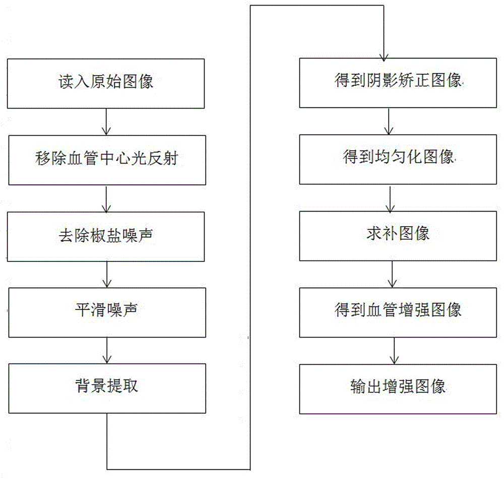
[0072] refer to figure 1 , a method for preprocessing retinal fundus images, comprising the steps of:

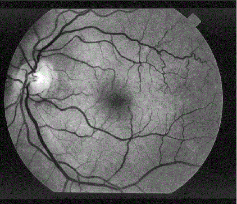
[0073] 1) Read in the original image: use the green channel to read in the original retinal fundus image, such as figure 2 Shown, retinal fundus

[0074] The original image is decomposed into red, green, and blue three-channel images, and the green channel with higher contrast is used for subsequent processing;

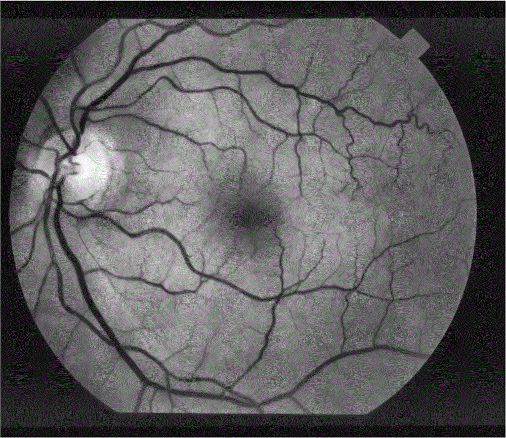
[0075] 2) Remove the light reflection of the blood vessel center: use mathematical morphology image filtering, such as image 3 As shown, the geometric features of the original retinal fundus image are extracted, and the square structural elements are selected according to the geometric features. The structural elements are simple and have a good expressive force on the geometric features of the object; Hit or not, you can get a morphological filter image that highlights the object characteristic information than the original retinal fundus image, remove the lig...


PUM


Abstract

The invention discloses a retinal fundus image preprocessing method. The retinal fundus image preprocessing method is characterized by including the steps of 1), reading an original image, namely reading the original image of the retinal fundus through a green channel; 2), removing blood vessel center light reflex; 3), removing salt-and-pepper noise; 4), smoothing noise; 5), conducting background extraction; 6), obtaining a shadow correction image; 7), obtaining a homogenization image; 8), solving a complementary image; 9), obtaining a blood vessel enhanced image; 10), outputting the enhanced image. The retinal fundus image preprocessing method has the advantages that the method enables the processed retinal image to be better in image quality and richer in information quantity, reserved enhancement of useful information such as blood vessels, optic disks and central foveae in the retinal image is achieved, and feature extraction, segmentation and recognition of the retinal image are improved.

Description

technical field [0001] The invention relates to feature extraction and image segmentation technology, in particular to a retinal fundus image preprocessing method. Background technique [0002] The arteries of the fundus retina are the only blood vessels that can be seen in the whole body of the human body. The blood vessels of the fundus can be observed intuitively, which reflects the blood vessels of the whole body. However, due to the influence of retinal vascular structure, imaging equipment, environment and noise, the quality of retinal images is generally poor. In order to facilitate the observation of retinal fundus, retinal images need to be preprocessed before use. [0003] Most of the current retinal vessel feature extraction and segmentation algorithms are based on enhanced retinal images, which requires that the processed retinal images should retain the original vessel texture information as much as possible, otherwise the final vessel segmentation accuracy will...

Claims


Application Information

Patent Timeline
IPC IPC(8): G06T7/00G06T7/11G06T5/00
CPCG06T7/0012G06T2207/20032G06T2207/30101G06T2207/30041G06T5/70
Inventor 王文涛何富运罗晓曙卢磊薛洋
Owner GUANGXI NORMAL UNIV
Learn More