Cancer immunotherapy using virus particles

A technology of virus particles and virus-like particles, applied in the direction of viruses, viral peptides, viruses/phages, etc.

Active Publication Date: 2017-08-01
CASE WESTERN RESERVE UNIV
1 Cites 1 Cited by

AI-Extracted Technical Summary

Problems solved by technology

The mechanistic basis for immune regulation by any VLP is...

Method used

[0034] In one aspect, the invention provides a method of treating cancer in a subject in need thereof by administering to the subject a therapeutically effective amount of a virus or virus-like particle. The virus or virus-like particle should be non-replicating and non-infectious in the subject to avoid infection of the subject. In some embodiments, the viral particles are administered in close proximity to the subject's tumor. Administering viral particles close to the tumor involves administering directly to the tumor site, thereby providing a high local concentration of viral particles in the tumor microenvironment.
[0095] The inventors sought to determine whether eCPMV therapeutic efficacy was limited to the B 16F10 metastatic lung model, or whether the immune-modulating antitumor effects were transferred to other models. The 4T1 BALB/c syngeneic breast cancer model, which is...

Abstract

A method of treating cancer in a subject in need thereof is described that includes administering a therapeutically effective amount of a virus or virus-like particle to the subject, wherein the virus particle is nonreplicating and noninfectious in the subject. The method represents a type of in situ vaccination, in which application of an immunostimulatory reagent directly to the tumor modifies the tumor microenvironment so that the immune system is able to respond to the tumor.

Application Domain

Organic active ingredientsSsRNA viruses positive-sense +5

Technology Topic

Wilms' tumorTumor microenvironment +4

Image

  • Cancer immunotherapy using virus particles
  • Cancer immunotherapy using virus particles
  • Cancer immunotherapy using virus particles

Examples

  • Experimental program(2)

Example Embodiment

[0059] Example 1: In situ vaccination with plant-derived viroid nanoparticle immunotherapy inhibits metastatic cancer
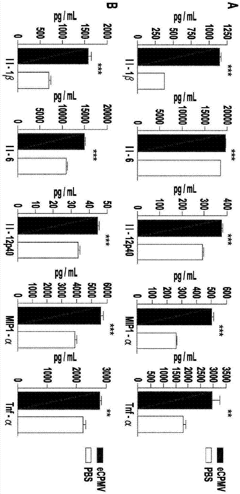
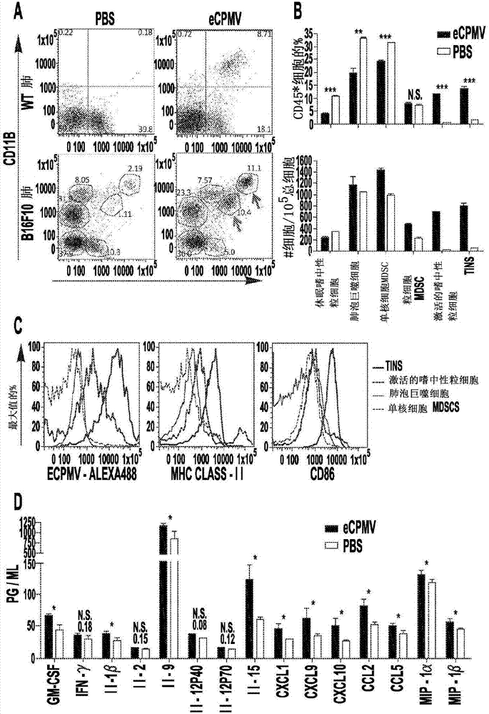
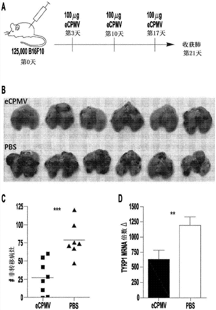
[0060] Current therapies are often ineffective against metastatic cancer, and emerging immunotherapies, while promising, are in the early stages of development. In situ vaccination refers to a process in which an immunostimulatory agent applied directly to the tumor alters the immunosuppressive microenvironment so that the immune system can respond efficiently to the tumor. The inventors hypothesized that treatment of lung tumor-bearing mice with viroid nanoparticles could modulate the immune environment of the lung and prevent metastatic injury of the B 16F10 class. This example shows that inhalation of self-assembled virus-like particles derived from cowpea mosaic virus (CPMV) inhibits tumor development in the lungs of mice following intravenous challenge. The inconsistency in tumor burden between CPMV- and PBS-treated mice was prominent and due to the presence of -/- , II-12 -/- or NOD/scid/II2Rγ -/- These phenomena were not seen in mice, so the effect is immune-mediated. In the absence of Ly6G + In the case of cells, it also loses potency. In the tumor microenvironment, CPMV nanoparticles are rapidly taken up by granulocytes, resulting in their potent activation and cytokine/chemokine production. CPMV nanoparticles are stable, non-toxic, highly scalable and can be modified with pharmaceuticals and antigens. These properties, combined with their inherent immunogenicity and remarkable potency against poorly immunogenic tumors, make CPMVs an attractive new immunotherapy against cancers that metastasize to the lung. In addition, CPMV has demonstrated clear therapeutic efficacy in a variety of other tumor models, including cutaneous melanoma, metastatic breast, colon, and ovarian cancer. This is the first report of viroid nanoparticles as cancer immunotherapy with proven therapeutic efficacy.
[0061] Materials and methods
[0062] Production and characterization of eCPMV
[0063] The eCPMV plasmid is produced by agroinfiltration of Nicotiana benthamiana plants using a culture of Agrobacterium tumefaciens LBA4404 transformed with the binary plasmid pEAQexpress-VP60-24K, wherein the binary plasmid comprises Genes for the capsid protein precursor VP60 and its 24K viral protease (thus cleaving it into the mature form). Six days after infiltration, leaves were harvested and eCPMVs were extracted using the established procedure. Using a UV/vis spectrometer (ε 280nm = 1.28mg -1 mL cm -1 ) to measure particle concentration, and to determine particle integrity by transmission electron microscopy and fast protein liquid chromatography.
[0064] mouse
[0065] C57BL/6J (01C55) female mice were purchased from the National Cancer Institute or The Jackson Laboratory. Il-12p35 -/- (002692), Ifn-γ -/- (002287), BALB/c (000651), and NOD/scid/IRy -/- (005557) female mice were purchased from The Jackson Laboratory. All animal studies were performed in accordance with the Institutional Animal Care and Use Committee of Dartmouth.
[0066] tumor model
[0067] The B16F10 murine melanoma cell line was obtained from Dr. David Mullins (Geisel School of Medicine at Dartmouth College, Hanover, New Hampshire). 4Tl-luciferase murine breast cancer cells were provided by Ashutosh Chilkoti (Duke University, Durham, NC). B 16F10, 4Tl-luc, and CT26 were grown in complete medium (RPMI, supplemented with 10% FBS and penicillin/streptomycin). ID8-Defb29/Vegf-A ovarian serous carcinoma cells in situ were cultured in complete medium supplemented with sodium pyruvate as previously described. Lizotte et al., Oncoimmunology, 3:e28926. eCollection 2014. Cells were harvested, washed in phosphate-buffered saline (PBS), and, depending on the tumor model, injected as follows: 1.25 x 10 in 200 μl PBS was injected intravenously in the rat tail vein. 5 Live cells (metastatic lung); 1.25xl0 in 30 μL PBS was injected intradermally in the right abdomen 5 Live cells (B16F10 flank); 1xl0 in 30 μL PBS was injected intradermally in the right part 5 Live cells (CT26 flank); intraperitoneal injection of 2xl0 in 200 μL PBS 6 Live cells (ID8-Defb29/Vegf-A peritoneal ovary). For 4Tl-luc tumor challenge, on day 0 will be 1xl0 in 30PBS 5 Live cells were injected into the left mammary fat pad, and the tumor was surgically removed on day 16, which was the day to which it was well established that the tumor spontaneously metastasized to the lung. Complete removal of the primary 4Tl-luc tumor was demonstrated by bioluminescent imaging. For the C57BL6J strain, B16F10 and ID8-Defb29/Vegf-A are homologous, while for the BALB/c background, CT26 and 4Tl-luc are homologous.
[0068] eCPMV Treatment Schedule
[0069] To WT challenged intravenously with B16F10, Il-12 -/- ,Ιfn-γ- -/- ,NOD/scid/IL2Rγ- -/- , and Ly6G-depleted mice were intubated and injected intraductally with 100 μg eCPMV in 50 μL PBS on days 3, 10 and 17 after tumor challenge. For the lung challenge assay, mice were euthanized on day 21 for quantification of metastatic-like lesions and tyrosinase expression. 4Tl-luc-bearing mice were injected intratracheally with 20 μg eCPMV in 50 μL PBS on day 16 (the same day as the primary tumor removal) and day 23 (7 days after tumor removal). Once the tumor reaches 10mm 2 , B16F10 flank tumors were injected intratumorally with 100 μg eCPMV on day 7 after tumor challenge and re-injected on day 14. Once the tumor reaches 10mm 2 , CT26 flank tumors were intratumorally injected with 100 μg eCPMV on day 8 after tumor challenge and re-injected on day 15. The diameter of the flank tumor was measured every other day, and when the tumor reached 200mm in diameter 2 , the mice were euthanized. Starting on day 7 after tumor challenge, ID8-Defb29/Vegf-A mice were injected IP with 100 μg eCPMV, and when tumors reached 35 g, mice were euthanized due to the development of ascites.
[0070] Antibodies and Flow Cytometry
[0071] Anti-mouse antibody against CD45 (30-F11), MHC-II (M5/114.15.2), CD86 (GL-1), CDl lb (Ml/70), F4/80 (BM8), and from Biolegend Ly6G (1A8) and CD16/CD32 (93) from eBioscience are specific. WT and B16F10 lung tumor-bearing mice were injected intratracheally with 100 μg of Alexa-488-labeled CPMV particles and euthanized 24 h later. Lungs were harvested and isolated into a suspension of single cells using the Miltenyi Mouse Lung Isolation Kit (Catalog #130-095-927). Red blood cells were removed using a lysis buffer of 150 mM NH4Cl, 10 mM KHCO3, and 0.5 mM EDTA. Flow cytometry was performed on a MACSQuant analyzer (Miltenyi). Data were analyzed using FlowJo software version 8.7.
[0072] Analysis of tyrosinase mRNA expression
[0073] Whole lungs were isolated and total RNA was extracted using the RNeasy kit (Qiagen, 74104). use iScript TM cDNA Synthesis Kit (Bio-Rad, 170-8891) was used to synthesize cDNA. Use iQ TM Green Supermix (Bio-Rad, 170-8882), the use concentration is the primer of 0.5 μ M, in CFX96 TM q-PCR was performed on a Real-Time PCR Detection System (Bio-Rad). Fold changes in mRNA transcripts were calculated with all samples normalized to mouse Gapdh using the AACT method.
[0074] Cytokine test
[0075] For in vivo cytokine data, a homogeneous mixture of total lungs was harvested from B16F10 lung tumor-bearing mice 24 h after inhalation of 100 μg eCPMV particles, which is day 8 post tumor challenge. For in vitro cytokine results, in 200 complete medium in 96-well round bottom plates with 20 μg eCPMV or PBS, at 1xl0 6 Cells/well culture Bone marrow-derived dendritic cells (BMDCs) and thioglycolate-stimulated peritoneal macrophages derived from C57BL6 mice. Cytokines were quantified using the mouse 32plex Luminex assay (MPXMCYTO70KPMX32, Millipore).
[0076] cell exhaustion
[0077] Mice were injected with a Ly6G-depleted mAb (clone no. 1A8) purchased from Bio-X-Cell (Catalog #BE0075-1) and administered IP at a dose of 500 μg, followed by eCPMV treatment one day later and then weekly during the survival assay once. Depletion of more than 95% of the target cell population in the lung was confirmed by flow cytometry.
[0078] IVIS imaging
[0079] Mice were injected IP with 150 mg/kg firefly D-luciferin (PerkinElmer #122796) in PBS and allowed to rest for 10 min. Imaging was performed using the Xenogen Vivo Vision IVIS Bioluminescent and Fluorescent Imager platform and analysis was performed using Living Image 4.3.1 software (PerkinElmer).
[0080] statistics
[0081] Unless otherwise stated, all experiments were repeated at least 2 times using 4-12 biological replicates and results were similar between replicates. Figures indicate statistical significance as p<0.05 for *, p<0.01 for **, and p<0.001 for ***. A p value <0.05 was considered statistically significant. Data for histograms were calculated using an unpaired Student's t-test. Error bars represent the standard error between the mean and the individual samples tested within the represented assay. Flank tumor growth curves were analyzed using two-way ANOVA. Generate tests using the log-rank Mantel-Cox test for survival analysis. Statistical analysis was performed using GraphPadPrism 4 software.
[0082] result
[0083] eCPMV nanoparticles are inherently immunogenic
[0084] Previously published work (using VLPs as a therapeutic approach for respiratory pathogen infections) used a number of systems and reported immunomodulatory capabilities to varying degrees. Furthermore, these studies usually focus on the immune response to the antigens contained in the VLPs, rather than on the inherent immunogenicity of the particles themselves, as described in VLPs. Bessa et al., Eur J Immunol. 38(1):114- 26 (2008) not explicitly shown. Furthermore, whether some VLPs are more stimulating than others is unknown. In view of this, the present inventors proposed the use of eCPMV (eCPMV refers to "empty" cowpea mosaic virus particles lacking RNA) as a novel immunotherapy, and they attempted to determine the inherent immunogenicity of eCPMV for the first time. eCPMV VLPs were added to in vitro cultures of bone marrow-derived dendritic cells (BMDCs) and primary macrophages harvested from C57BL6 mice. After 24 hours of culture with eCPMV particles, induced BMDC ( figure 1 A) and macrophages ( figure 1 B) Secretion of higher levels of typical pro-inflammatory cytokines, including II-β, 11-6, Il-12p40, Ccl3 (MIP1-a) and Tnf-a, leading the inventors to conclude that eCPMV is inherently immunostimulatory .
[0085] eCPMV inhalation radically alters the microenvironment of B16F10 lung tumors
[0086] Next, the immunomodulatory effect of eCPMV inhalation on the lung microenvironment was determined in terms of changes in immune cell composition and cytokine and chemokine levels. Exposure of lungs of tumor-free mice to eCPMV showed that Ly6G + Neutrophils were significantly activated by the CD1 lb activation marker (Costantini et al., Int Immunol., 22(10):827-38(2010)) ( figure 2 A upper panel) and up-regulation of CD86 co-stimulatory markers were assessed. The Alexa488-labeling of the particles allowed for cell tracking, which enabled the inventors to confirm that it was this CD1lb + Ly6G + A subpopulation of activated neutrophils specifically takes up eCPMV.
[0087] The lungs of mice bearing B16F10 melanoma tumors showed a more complex composition of immune cells. By day 7, immunosuppressive CD11b was observed + Ly6G - F4/80 lo Class II - SSC lo Mononuclear myeloid-derived suppressor cells (MDSCs) and CD11b + Ly6G + F4/80 - Class II mid SSC hi A large population of granulocytic MDSCs ( figure 2 A bottom panel). Gabrilovich et al., Nat Rev Immunol., (4):253-68 (2012). The inventors also observed that the presence of CD11b + Ly6G + Class II mid CD86 hi A small population of cells, which have been described in the literature as "tumour-infiltrating neutrophils" or "N1 neutrophils," are known to respond through a coordinated adaptive immune response, high levels of pro-inflammatory The production of cytokines, the recruitment of T and NK cells, and the direct cytotoxic effect on tumor cells play an anti-tumor role. Fridlender et al., Cancer Cell. 16(3):183-94(2009); Mantovani et al, Nat Rev Immunol.(8):519-31(2011). Inhalation of eCPMV into B16F10-bearing lungs drastically altered immune cell composition 24 hours after administration. Tumor-infiltrating neutrophils (TIN) and CD11b were also observed + Ly6G + Significant increase in activated neutrophil population, and CD11b - Ly6G + Decrease in dormant neutrophils ( figure 2 A bottom panel, see arrow). as CD45 + Percentage of cells as well as percent of total number, TIN and activated CD11b + A sharp increase in the neutrophil population ( figure 2 B). Interestingly, it is these subpopulations of neutrophils that account for an extremely large number of eCPMV particles, especially TIN, which is much higher than CD11b + Activated neutrophils occupy 10 times more eCPMV ( figure 2 C). Monocyte MDSC, resting neutrophil and alveolar macrophage populations did not occupy eCPMV, whereas granulocyte MDSC displayed non-inhibited uptake. TINs and activated neutrophil populations also express MHC class II, and in particular, TINs display high levels of the co-stimulatory marker CD86, suggesting potential antigen presentation and T cell priming capabilities. No significant changes were observed in a large number of monocytes or granulocytes MDSCs.
[0088] Activation of neutrophil populations by eCPMV is consistent with data collected from multi-throughput cytokine/chemokine arrays on a homogeneous mixture of whole lungs (treated with eCPMV or PBS) of B16F10 tumor-bearing lungs Implementation( figure 2 D). Specifically, marked increases in the neutrophil chemoattractants GM-CSF, Cxcll, Ccl5, and MIP-Iα were seen, as well as cytokines and chemokines produced by activated neutrophils (e.g., GM-CSF, II -1β, 11-9, Cxcll, Cxcl9, CxcllO, Ccl2, MIP-la and MIP-Ιβ) were significantly increased. Interestingly, the inventors did not observe a significant increase in the levels of 11-6 or Tnf-a, which are traditional pro-inflammatory cytokines, which are disadvantageous in lung immunobiology.
[0089] eCPMV inhalation inhibits the development of metastatic lung tumors of the B16F10 class
[0090] Next, the inventors investigated whether the inherent immunogenicity of eCPMV particles in the lung induced anti-tumor immunity in the B16F10 intravenous model of invasive metastatic carcinoma. In fact, weekly intratracheal injections of 100 μg eCPMV ( image 3 A) significantly reduced tumor burden, which can be measured by the number of metastatic tumor lesions ( image 3 , B and C) and tyrosinase ( image 3 D) expression to evaluate. Tyrosinase-related protein 1 (Tyrp1) is a melanocyte-specific gene (Zhu et al., Cancer Res., 73(7):2104-16(2013)), and its expression in the lung is restricted to B16F10 tumors cells, which can quantitatively measure weight development and serve as a control for size-altered metastatic-like lesions.
[0091] The antitumor efficacy of eCPMV inhalation is immune-mediated
[0092] The inventors determined that in the following transgenic mice: Il-12 -/- , Ifn-γ -/- , and NOD/scid/IL2Rγ -/- in, by repeating image 3 Whether the experimental design indicated requires immune cells for therapeutic efficacy. In the absence of II-12 ( Figure 4 A), Ifn-γ( Figure 4 B), or in the absence of T, B and NK cells ( Figure 4 C) In NSG mice, no significant difference in lung tumor burden between eCPMV- and PBS-treated mice was observed.
[0093] In the lungs of mice bearing B16F10 tumors, eCPMV accumulated in neutrophils and activated the cells ( figure 2 , A to C), in addition, significant increases in neutrophil-associated cytokines and chemokines were detected in the lungs of mice after eCPMV inhalation ( figure 2 D). Therefore, the use of monoclonal antibodies to deplete neutrophils is necessary to assess the efficacy of neutrophils for treatment. Depleting neutrophils from the lungs of tumor-bearing mice abolished the antitumor effect we observed in WT mice ( Figure 4 D). This situation, combined with the Il-12 -/- , Ifn-γ -/- , and the lack of potency observed in NSG mice, led the inventors to infer that the antitumor effect of eCPMV inhalation on B16F10 development is through immune regulation of the lung tumor microenvironment, specifically requiring the neutrophil compartment ( compartment) exists.
[0094] eCPMV antitumor potency is not limited to the B16F10 intravenous lung model
[0095] The inventors sought to determine whether eCPMV therapeutic efficacy was restricted to the B 16F10 metastatic lung model, or whether the immunomodulatory antitumor effects were transferred to other models. The 4T1 BALB/c syngeneic breast cancer model, which is metastatic and truly metastatic, was first used. By day 16, 4T1 tumors established in the mammary fat pad spontaneously metastasized into the lungs, at which point the primary tumors were surgically removed and eCPMV therapy was initiated, in which intratracheal injections were performed, thereby affecting the lung tumors. develop. 4T1 cells also express luciferase, allowing the inventors to follow the development of metastatic lung tumors. Mice treated intratracheally with eCPMV particles had significantly prolonged lung tumor onset and significantly prolonged survival ( Figure 5 A). Mice from the eCPMV and PBS treated groups had comparable primary mammary fat pad tumor burden on the day of surgical removal. Thus, differences in tumor development and survival were due to the treatments described. No mice from either group experienced recurrence of primary mammary fat pad tumors.
[0096] To determine whether the robust efficacy of eCPMV particles in B16F10 and 4T1 metastatic lung models is unique to the lung immune context, their ability to treat skin tumors was also examined. After direct injection of eCPMV, intradermal B16F10 melanoma ( Figure 5 B) and CT26 colon ( Figure 5 C) The growth of tumors was significantly delayed, and half of the B16F10eCPMV-treated mice eliminated tumors together after only 2 treatments ( Figure 5 B).
[0097] Finally, the therapeutic effect of eCPMV was investigated in a disseminated peritoneal serous ovarian cancer model. Conejo-Garcia et al., Nat Med., (9):950-8 (2004). Weekly eCPMV-treated Defb29/Vegf-A-challenged mice exhibited significantly improved generation relative to PBS-treated controls ( Figure 5 D). In fact, mice receiving eCPMV outlived other immunotherapies tried in this model, including live attenuated strains of Listeria monocytogene (Lizotte et al., Oncoimmunology , 3:e28926.eCollection 2014), the avirulent Toxoplasma gondii (Baird et al., Cancer Res., 73(13):3842-51(2013)), the CD40 and poly( I:C) (Scarlett et al., Cancer Res.,69(18):7329-37(2009)), or 11-10 blocking antibody (Hart et al., T Cell Biol.,2:29(2011) ). It is important to note that since in vitro exposure to high concentrations of particles had no effect on the viability or proliferation of cancer cells, the antitumor effect of eCPMV in the models examined did not help direct the cytotoxicity of tumor cells, the logical conclusion being that The striking antitumor effects induced by eCPMV nanoparticle treatment were fully immune-mediated and could be translated into a variety of tumor models at diverse anatomical sites.
[0098] discuss
[0099] eCPMV nanoparticles are immunotherapeutics of surprising heights and clearly modulate the tumor immune environment. BMDCs and macrophages exposed to the particles robustly secreted selective pro-inflammatory cytokines ( figure 1 , A and B). The eCPMV particles are produced in plants, then polyvinylpyrrolidone is used to extract plant contaminants, eCPMV is separated by PEG precipitation and sucrose gradient centrifugation, and finally the particles are concentrated by ultrapelleting. Purity was checked by UV/Vis absorbance, transmission electron microscopy (TEM), fast protein liquid chromatography (FPLC), and SDS gel electrophoresis. Wen et al., Biomacromolecules, 13(12):3990-4001 (2012). No contaminants were detected during these processes. The particles were then suspended in PBS. Furthermore, eCPMV completely lacks RNAs that can stimulate TLR3/7. Therefore, the inventors concluded that the high immunogenicity of eCPMV is due to the size, nature or innate immune recognition of the viral capsid protein. This is in contrast to viroid or protein cage nanoparticles produced in E.coli or other systems, which can contain immunogenic contaminants such as endotoxin or viral nucleic acids, which interfere with migration during purification. Except challenging.
[0100] eCPMV inhalation alters the lung tumor microenvironment and requires Ly6G + Neutrophils, the population least correlated with response to tumor immunotherapy, are present. Notably, eCPMV particles appear to specifically target Ly6G + neutrophils. eCPMV has been shown to bind to surface vimentin (Steinmetz et al., Nanomed.6(2): 351-64 (2011)), but it is unknown whether lung remnant neutrophils internalize the granules by this mechanism, or whether surface expression of vimentin is characteristic of murine neutrophils. eCPMV was shown to target dormant neutrophils and convert them into activated CD11b + Phenotype and CD11b + -Class II + CD86 hl Tumor-infiltrating neutrophils. In fact, these two populations (activated neutrophils and tumor-infiltrating or "N1" neutrophils) were the only populations of innate immune cells that changed significantly and rapidly following eCPMV lung exposure, which dramatically increased CD45 + The percentage of cells and the percentage of total cell number ( figure 2 B). Neutrophils are canonically regarded as sentinels of microbial infection, rapidly engulfing and killing bacteria followed by apoptosis, but they have an emerging role in tumor immunology. Although still controversial, neutrophils have been shown to have phenotypic plasticity in M1/M2 polarization similar to that accepted in the macrophage literature. Studies have shown the infiltration of an immunosuppressive pro-angiogenic "tumor-associated neutrophil" population in B16F10 metastatic lung cancer, human liver cancer, sarcoma and lung adenocarcinoma models (associated with enhanced tumor progression). Alternatively, depleting tumor residual immunosuppressive neutrophils, converting them to a pro-inflammatory phenotype, or recruiting activated neutrophils to infiltrate the tumor microenvironment is associated with therapeutic efficacy. Activated neutrophils can directly kill tumor cells by releasing reactive oxygen species intermediates (ROI), inducing (prime) CD4 + T cells, and polarize them to a Thl phenotype, cross-prime CD8 + T cells, and regulate NK cell survival, proliferation, cytotoxic activity and IFN-γ production. Activated neutrophils can also produce the Cxcr3 ligands Cxc19 and Cxc110, which can recruit CD4 associated with antitumor immunotherapy efficacy in melanoma models + and CD8 + T cells. Data showing an increase in the immunostimulatory neutrophil population ( figure 2 B) Data consistent with cytokines ( figure 2 D) in which an increase in the neutrophil chemoattractants GM-CSF, Cxcll, Ccl5, MIP-Iα, cytokines and chemokines known to be produced by neutrophils was observed, including GM-CSF, III - Iβ, 11-9, Cxcll, Cxcl9, CxcllO, Ccl2, MIP-la and MIP-1β. This data is correspondingly in line with in vivo tumor progression data showing that neutrophils are required for eCPMV antitumor efficacy. Interestingly, although the levels of many cytokines and chemokines increased to statistically significant levels after eCPMV treatment, the changes were most modest compared to the large differences in actual tumor burden ( image 3 , B to D). Furthermore, no increase was observed in the pro-inflammatory cytokines Tnf-a or 11-6, which are known to cause tissue damage when upregulated in the lung. Thus, it was shown that eCPMV treatment of lung tumors is effective and does not elicit such an inflammatory cytokine response leading to acute non-injury.
[0101] eCPMV inhalation showed significant efficacy as a monotherapy ( image 3 ), the monotherapy is most clearly immune-mediated ( Figure 4 ). Since eCPMV particles do not directly kill tumor cells, or share any antigenic overlap with B 16F10 tumors, but would elicit an antitumor response (which requires Th1-associated cytokines 11-12( Figure 4 A) and Ifn-γ( Figure 4 B), adaptive immunity ( Figure 4 C) and neutrophils ( Figure 4 D)), so the method described is novel. This suggests that when eCPMV is introduced into the lung, the intrinsic immunogenicity of eCPMV disrupts the tolerogenic nature of the tumor microenvironment; thereby, in essence, removing the hindrance to the existing suppressed anti-tumor immune response, Or allow the development of new anti-tumor responses.
[0102] This work also shows that the eCPMV antitumor efficacy in the intravenous B 16F10 metastatic lung model is not an artifact of the C57BL6 mouse strain or the B16F10 model, as eCPMV therapy was effective in flank B 16F10, ovarian, and metastatic breast and colon cancers. Impressively equivalent in the 2 BALB/c models of ( Figure 5 ). Constitutive luciferase expression and intradermal challenge of B16F10 and CT26 in 4T1 breast cancer cells allowed us to quantitatively measure tumor progression in a manner not available for the B16F10 lung model. It is in these models that we observe potent and immediate antitumor effects that significantly delay tumor progression and, in the case of B 16F10 and CT26 intradermal tumors, cause rapid involution of established tumors and necrotic center formation ( Figure 5 , B and C) (which remained localized within the margins of the tumor and did not appear to affect surrounding tissue). This early response to eCPMV at day 3 after intratumoral injection suggests that eCPMV particles induce innate immune cell-mediated antitumor responses. However, subsequent studies to determine the infiltration of immune cells in these tumors and the phenotype of such cells are intriguing.
[0103] This is the first report of VLP-based nanoparticles as cancer immunotherapy and further immunotherapy that specifically targets and activates neutrophils in the tumor microenvironment to coordinate downstream anti-tumor immune responses. eCPMV nanoparticles alone are immunogenic and highly effective as monotherapy. However, it can also be used as a nanocarrier for tumor antigens, pharmaceuticals or immune adjuvants, opening the exciting possibility that eCPMV can be modified to deliver a payload that further enhances and improves its immunotherapeutic efficacy.
[0104] The inventors demonstrated that eCPMV is an effective immunotherapy against tumors originating in various organs. eCPMV therapy was well tolerated at doses comparable to other biological and small molecules, and had no observed toxic side effects. In conclusion, eCPMV self-assembled VLPs are an exciting new platform for cancer therapy that exerts its therapeutic efficacy by unleashing the power of anti-tumor immunity.

Example Embodiment

[0105] Example 2: eCPMV Treatment of Skin B16F10 Induces Systemic Anti-Tumor Immunity
[0106] like Image 6 As shown, the inventors established skin melanoma tumors, and injected eCPMV particles directly into the skin (100 μg/injection, arrows indicate injection days). The above treatment resulted in the complete disappearance of tumors in half of the mice. In cured mice, we then waited 4 weeks and challenged again with the same tumor cells, but we injected tumor cells on the opposite flank. The majority of these mice did not develop secondary tumors. For clarity, primary tumors were injected directly with particles, while mice bearing secondary tumors were left untreated. They must rely on a separate systemic antitumor immune memory. eCPMV was applied locally in the primary tumor, but systemic immunity was induced. "Re-challenged" mice that previously had B 16F10 skin tumors were injected directly, and shrunk and disappeared.

PUM

no PUM

Description & Claims & Application Information

We can also present the details of the Description, Claims and Application information to help users get a comprehensive understanding of the technical details of the patent, such as background art, summary of invention, brief description of drawings, description of embodiments, and other original content. On the other hand, users can also determine the specific scope of protection of the technology through the list of claims; as well as understand the changes in the life cycle of the technology with the presentation of the patent timeline. Login to view more.
Why Eureka
Try Eureka