

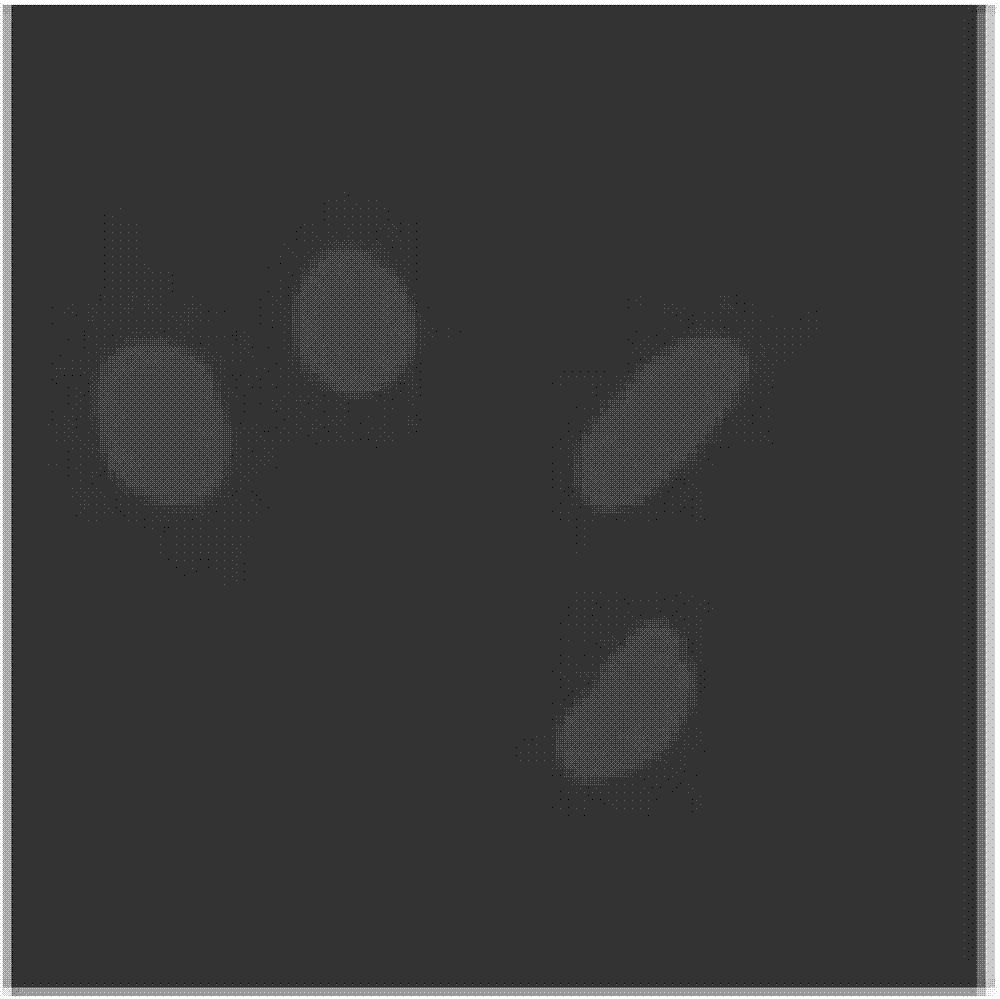
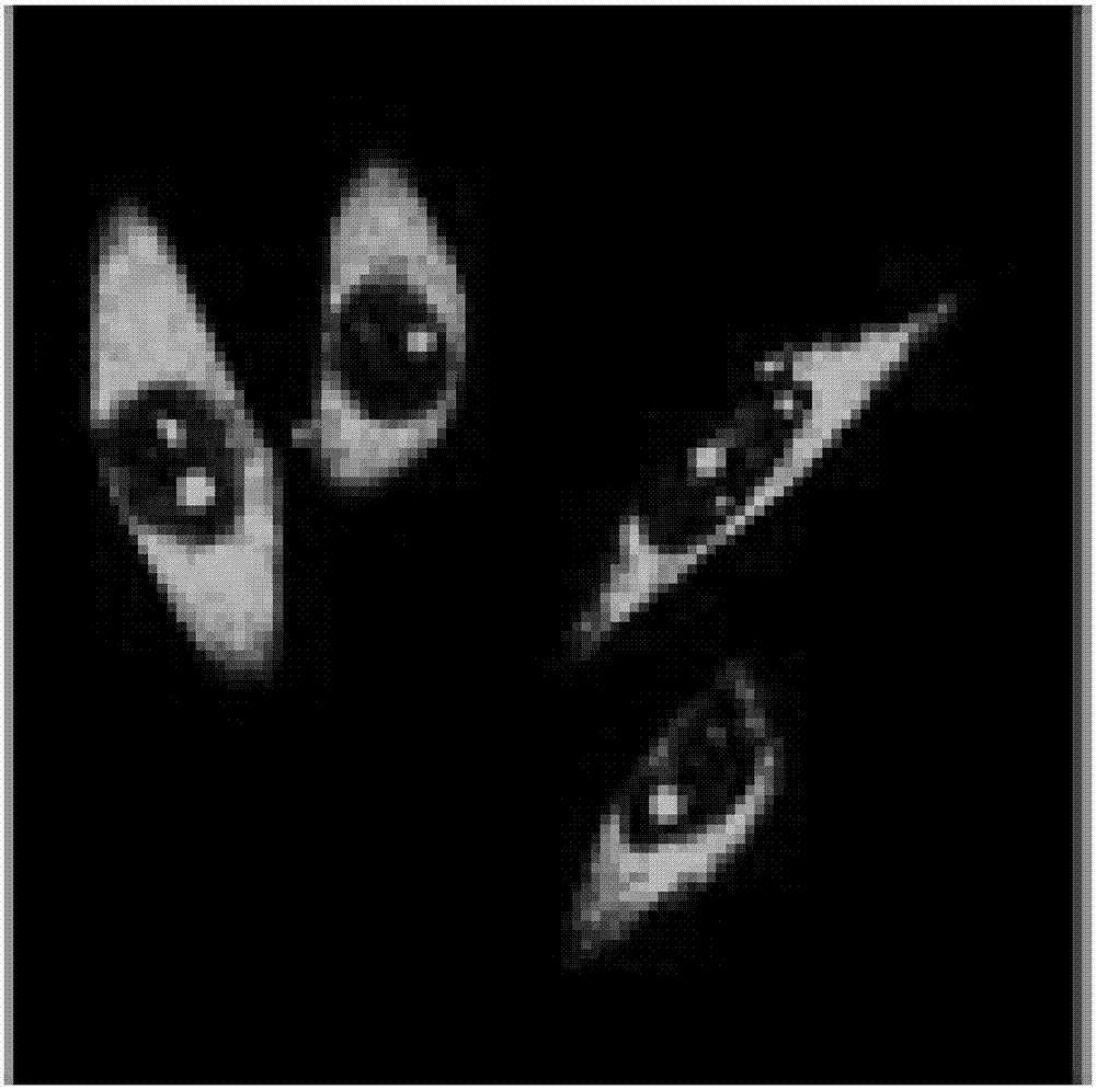
Fluorescent probes for simultaneously displaying cell nucleus structure and cell integral morphology in live cells
A fluorescent probe and simultaneous display technology, applied in the field of fluorescent probes, can solve the problems of fluorescent reagents limiting biological research, disease diagnosis, pathological research and drug development, etc., and achieve strong photostability, good counterstaining compatibility, strong color effect
- Summary
- Abstract
- Description
- Claims
- Application Information
AI Technical Summary
Problems solved by technology
Method used
Image


Examples
Embodiment 1
[0032] Example 1: Probe preparation.
[0033] 2-(4-ethoxyphenyl)-6-(4-methyl-1-piperazinyl)-1H,3H-2,5-dibenzimidazole-trihydrochloride, 1,1'-bis Octadecyl-3,3,3',3'-tetramethylindolocarbocyanine perchlorate was purchased from Molecular Probes Inc., and the article numbers were Hoechst33342 and DiI, respectively.
[0034] (E)-1-(2-hydroxyethyl)-4-(2-(1-methyl-1H-pyrrole-2-)vinyl)pyridinium iodide salt according to the patent “a kind of Synthesized by the experimental process described in "Pyrrole pyridinium salt fluorescent probe for RNA and nucleolus imaging in living cells (application number: 201310215970.9)".
Embodiment 2
[0035] Example 2: SiHa cell culture.
[0036] Adherently culture SiHa cells in culture medium containing 10% fetal bovine serum at 37°C, 5% CO 2 Cultured in a saturated humidity incubator, and subcultured once every 2-3 days. When the cells grow to the logarithmic phase, culture the slices: ① Soak the coverslips in absolute ethanol for 30 minutes, dry them with an alcohol lamp and place them in a disposable 35mm culture dish for later use; Wash the cells three times with PBS, digest with 1mL 0.25% trypsin for 3-5 minutes, pour out the trypsin carefully, add fresh culture medium and pipette evenly, and count the cells. The concentration is 1×10 5 , and then inoculated into the above-mentioned petri dishes containing coverslips, and cultured in a 5% CO2 incubator to allow the cells to grow on the sheets. After the SiHa cells grow on the slide and cover the glass, it is used for the experiment.
Embodiment 3
[0037] Example 3: (E)-1-(2-hydroxyethyl)-4-(2-(1-methyl-1H-pyrrole-2-)vinyl)pyridinium iodide salt, 2-(4-ethoxy Phenyl)-6-(4-methyl-1-piperazinyl)-1H,3H-2,5-dibenzimidazole-trihydrochloride, 1,1'-octadecyl-3, 3,3',3'-Tetramethyldocarbocyanine perchlorate cell counterstaining method:
[0038] First prepare 5mM (E)-1-(2-hydroxyethyl)-4-(2-(1-methyl-1H-pyrrole-2-)vinyl)pyridinium iodide DMSO solution, 1mM 2-( 4-ethoxyphenyl)-6-(4-methyl-1-piperazinyl)-1H,3H-2,5-dibenzimidazole-trihydrochloride DMSO solution, 1mM 1,1'- Octacosyl-3,3,3',3'-tetramethyldocarbocyanine perchlorate DMSO solution was used as the mother liquor.
[0039] Take 4 μL of (E)-1-(2-hydroxyethyl)-4-(2-(1-methyl-1H-pyrrole-2-)vinyl)pyridinium iodide mother solution, 5 μL of 2-(4- Ethoxyphenyl)-6-(4-methyl-1-piperazinyl)-1H,3H-2,5-dibenzimidazole-trihydrochloride stock solution and 5 μL of 1,1'-octacos Alkyl-3,3,3',3'-tetramethyldocarbocyanine perchlorate mother solution was added to 1 mL of PBS buffer and mixe...
PUM
| Property | Measurement | Unit |
|---|---|---|
| diameter | aaaaa | aaaaa |
Abstract
Description
Claims
Application Information