X-ray chest radiograph bone suppression processing method based on wavelet decomposition and convolutional neural network

A convolutional neural network and wavelet decomposition technology, applied in the field of X-ray chest radiograph bone suppression processing, can solve the problems of anatomical structure image overlap, reduce radiation dose and motion artifacts, and improve prediction accuracy.

Inactive Publication Date: 2017-08-11
SOUTHERN MEDICAL UNIVERSITY
View PDF3 Cites 25 Cited by
  • Summary
  • Abstract
  • Description
  • Claims
  • Application Information

AI Technical Summary

Problems solved by technology

[0006] The purpose of the present invention is to avoid the deficiencies of the prior art and provide a X-ray thoracic bone suppression processing method based on wavelet decomposition and conv

Method used


Image

  • X-ray chest radiograph bone suppression processing method based on wavelet decomposition and convolutional neural network
  • X-ray chest radiograph bone suppression processing method based on wavelet decomposition and convolutional neural network
  • X-ray chest radiograph bone suppression processing method based on wavelet decomposition and convolutional neural network

Examples


Embodiment 1

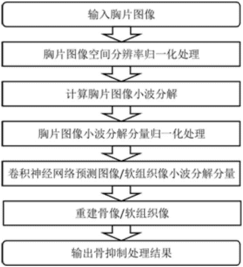
[0044] The X-ray chest X-ray bone suppression processing method based on wavelet decomposition and convolutional neural network comprises the following steps: step (1), normalization processing of X-ray chest X-ray image spatial resolution; Step (2), obtaining X-ray Chest film image wavelet coefficient; Step (3), the normalization process of chest film image wavelet coefficient; Step (4), the increase of training sample and the deletion of artifact area, and its training sample sampling preprocessing; Step (5 ), training the convolutional neural network for predicting bone images or soft tissue image wavelet coefficients; step (6), reconstructing bone images or soft tissue images through predicted wavelet coefficient images; step (7), in the chest image after the original normalization Subtract the reconstructed bone image or use the reconstructed soft tissue image as the result of bone suppression. Such as figure 1 Shown is the basic flow chart of the present invention for b...

Embodiment 2

[0078] Based on wavelet decomposition and convolutional neural network X-ray thoracic bone suppression processing method, other structures are the same as in Example 1, the difference is:

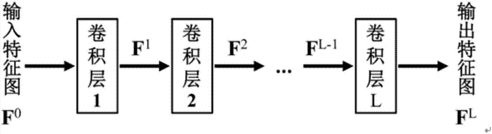
[0079] The output of the convolutional neural network prediction is the wavelet coefficient image of the bone image, and the spatial size corresponding to the pixel size of the input image is normalized to 0.194mm; the convolutional neural network contains three convolutional layers in each prediction unit, the first layer The convolution kernel size of the convolutional layer is 16×16, the number of convolution kernels is 256, the convolution kernel size of the second convolution layer is 1×1, and the number of kernels is 256, and the convolution kernel size of the third convolution layer is is 8×8, and the number of cores is 256; the nonlinear activation function after the first and second convolutional layers is the ReLU function. The haar wavelet is used to decompose the chest image wav...

Embodiment 3

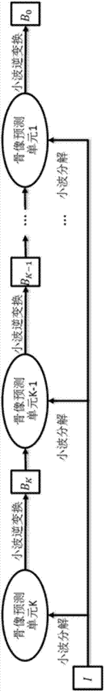
[0082] The X-ray chest X-ray bone suppression processing method based on wavelet decomposition and convolutional neural network, the other structures are the same as in Example 1, the difference is that the bone suppression processing is performed through a multi-scale cascade method, and the bone image in the multi-scale cascade framework The number of prediction units is 4, the wavelet decomposition series of the bone image prediction unit k to the input image is 24-k, and the wavelet inverse transform B of the predicted output component k The scale of is 2 times of that before prediction. The pixel size of the input chest image was normalized to 0.194mm. The convolutional neural network in the bone image prediction unit k contains three convolutional layers. The first layer has a convolution kernel size of 16×16 and the number of kernels is 256, and the second layer has a convolution kernel size of 1×1 and the number of kernels is 256, the size of the convolution kernel of...


PUM


Abstract

The invention discloses an X-ray chest radiograph bone suppression processing method based on wavelet decomposition and a convolutional neural network. By adopting a convolutional neural network structure and using a chest radiograph image wavelet coefficient as the input, a wavelet coefficient image of a corresponding bone image or soft tissue image is predicted. The existing bone image or soft tissue image artificial neural network prediction method processes an original chest radiograph image by adopting a relatively complex contrast normalization method, whereas this method processes the input chest radiograph image in a wavelet domain, and can normalize the amplitude by adopting a simple method; and the existing bone image or soft tissue image artificial neural network prediction method needs to design an image feature extraction method as the input of the artificial neural network, whereas this method completes an image feature extraction process by directly using the wavelet decomposition image of the chest radiograph image as an input, training the convolutional neural network to learn automatically and optimizing the convolution kernel, so the image feature extraction method does not need to be designed.

Description

technical field [0001] The invention relates to the technical field of digital image processing, in particular to an X-ray chest X-ray bone suppression processing method based on wavelet decomposition and convolutional neural network. Background technique [0002] Chest X-ray plain film (abbreviated as chest film) is one of the basic imaging methods for lung disease detection. However, the overlapping of anatomical structures in chest radiographs increases the difficulty for doctors to read and diagnose, especially the occlusion of ribs and clavicles will make the diagnosis of small pulmonary nodules more difficult. [0003] The method that prior art solves the above problem mainly contains following two major directions: one, along with the development of equipment and technology of digital X-ray imaging (Digital Radiography, DR) and computer X-ray photography (Computed Radiography, CR), X-ray dual Dual Energy Subtraction (DES) technology can separate images showing charac...

Claims


Application Information

Patent Timeline
IPC IPC(8): G06T5/00
CPCG06T5/003G06T2207/10116G06T2207/20064G06T2207/20081G06T2207/20084
Inventor 阳维陈莹胤贠照强冯前进
Owner SOUTHERN MEDICAL UNIVERSITY
Why Eureka
Try Eureka