Blood vessel closure structure which can be completely degraded

A blood vessel closure and blood vessel technology, applied in the field of medical devices, can solve the problems of occupying medical staff's time, long-term bed rest of patients, vagus nerve reflex, etc., and achieve the effect of shortening bed rest time, simplifying operation steps, and reducing burden

Inactive Publication Date: 2018-03-16
LEPU MEDICAL TECH (BEIJING) CO LTD
View PDF6 Cites 9 Cited by
  • Summary
  • Abstract
  • Description
  • Claims
  • Application Information

AI Technical Summary

Problems solved by technology

[0005] The technical problem to be solved by the present invention is the vagus nerve reflex existing in the traditional manual pressing method, the hemostasis time is long, the time of the medical staff is taken up, the patient needs to be bedridden for a long time, etc.; The suture removal problem brought about, the vascular closure structure is simplified, and the cost of learning and using is reduced

Method used


Image

  • Blood vessel closure structure which can be completely degraded
  • Blood vessel closure structure which can be completely degraded
  • Blood vessel closure structure which can be completely degraded

Examples


no. 1 example

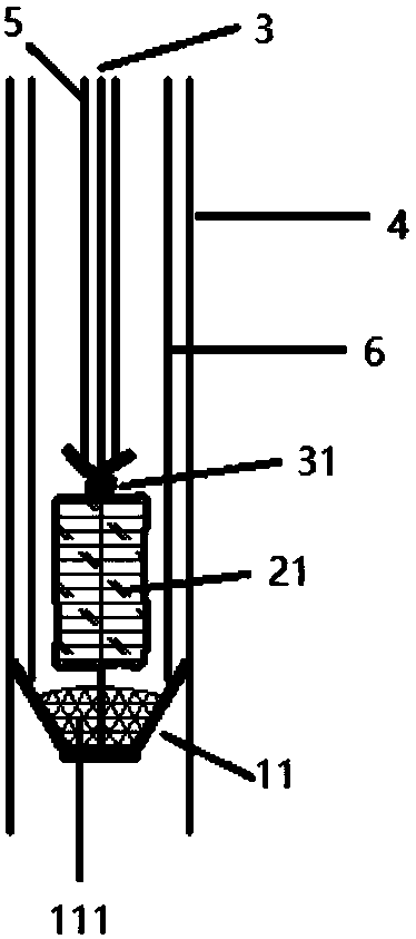
[0041] The fully degradable vascular closure structure provided by this embodiment is as figure 1 , 2 shown.

[0042] The blocking umbrella anchor 11 provided in this embodiment is made of a completely degradable material, which is made by laser cutting a degradable pipe and then heat-shaped; preferably, the surface of the blocking umbrella anchor 11 is covered with a 111 flow blocking film, and the The end is connected with suture 3. Firstly, the delivery sheath 4 is inserted into the vascular lumen along the puncture port, and then the push rod 6 is pushed to spread the occlusion umbrella anchor 11 in the lumen, the delivery sheath 4 is withdrawn, and then the suture 3 is pulled back to seal it. The blocking umbrella is anchored on the inner wall of the blood vessel; the presence of the blocking membrane 111 enables the structure to have a hemostatic effect during the anchoring process; preferably, the material of the blocking umbrella anchor 11 is PLA, and the material of...

no. 2 example

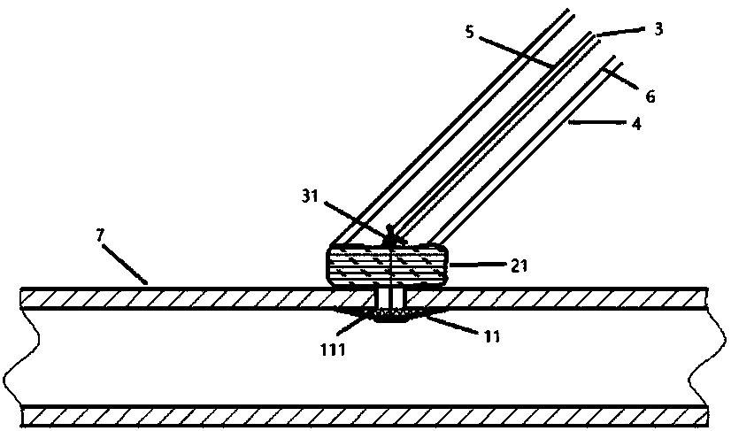
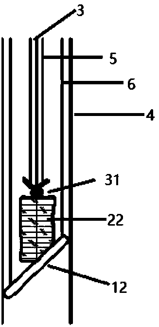
[0047] The fully degradable vascular closure structure provided by this embodiment is as image 3 , 4 shown.

[0048] The strip anchor 12 provided in this embodiment is made of completely degradable material; the strip anchor 12 is connected with the suture 3 . First, insert the delivery sheath 4 into the vessel cavity along the puncture port, then push the push rod 6 to release the strip anchor 12 in the lumen, withdraw the delivery sheath 4, and then pull the suture 3 back to make the strip anchor The anchor 12 is anchored on the inner wall of the blood vessel; preferably, the material of the strip anchor 12 is PGA.

[0049] After the bar-shaped anchor 12 is anchored, the push rod 6 is retracted until the bovine collagen tampon oppressor 22 is completely released on the outer wall of the lumen, such as Figure 4 Shown; Preferably, the constrictor material provided in this embodiment is bovine collagen, which is in the shape of a porous sponge and can be shrunk in the deli...

no. 3 example

[0053] The fully degradable vascular closure structure provided by this embodiment is as Figure 5 , 6 shown.

[0054] The balloon anchor 13 provided in this embodiment is a temporary anchor; preferably, the balloon anchor 13 is a spherical balloon made of Pebax. First, insert the delivery sheath 4 into the lumen of the blood vessel along the puncture port, then push the balloon inflation tube 8 to the marked point, at this time, the balloon is inflated, and then the balloon is retracted so that the balloon is close to the inner wall of the blood vessel to complete the anchoring Certainly.

[0055] After the temporary balloon anchor 13 is anchored, the delivery sheath 4 is retracted to the outer wall of the lumen, and the hydrogel (PEG hydrogel oppressor 23) is slowly injected along the injection tube 9, such as Figure 6 Shown; Preferably, the compressive material hydrogel material provided in this embodiment is PEG, which will absorb water and swell after being released o...


PUM


Abstract

The invention provides a blood vessel closure structure which can be completely degraded. The structure especially aims at arterial puncture mouth closure. The structure comprises an anchor in a bloodvessel lumen, an anti-hemorrhagic compression object outside the lumen and a connecting suture; the portions, kept in the body, of the structure are all made from material which can be completely degraded; according to one particularly preferable embodiment, the anchor in the blood vessel lumen is closely attached to the blood vessel inner wall and anchors a puncture mouth; the anti-hemorrhagic compression object presses the blood vessel outer wall tightly and blocks the puncture mouth; the suture connects the anchor and the compression object, and through an auto-tightened knot, the sandwichstructure is fixed. By means of the blood vessel closure structure which can be completely degraded, the burden of longstanding manual pressing for hemostasis of a doctor is reduced, and time in bedof a patient is shortened; compared with a blood vessel suture closure structure, the blood vessel blocking closure structure has the advantages that the operative steps are simplified, and the learning cost and the hemostasis failure rate of a surgeon are reduced; the objects implanted in the body can be completely degraded, so that a suture removing process brought by use of non-degradable material is avoided.

Description

technical field [0001] The invention relates to the technical field of medical devices, in particular to a fully degradable blood vessel closure structure. Background technique [0002] Vascular puncture is a necessary approach for interventional surgery, and manual hemostasis has always been the only option for operators. This method requires the operator to continuously apply near-blocking pressure to the vascular puncture point for 10-20 minutes, and strictly brake for 6-24 hours. However, there are vagus nerve reflexes, long hemostasis time, occupation of medical personnel's time, long-term bed rest and other shortcomings in the manual pressure hemostasis method. In recent years, devices targeting suturing at vascular puncture sites have emerged. The vascular suture device uses multiple sutures to tighten and knot the vascular puncture under the skin. Although it can effectively reduce hemostasis and nursing time, because it uses non-degradable sutures, the suture remo...

Claims


Application Information

Patent Timeline
Patent Type & Authority Applications(China)
IPC IPC(8): A61B17/12A61B17/06
CPCA61B17/06166A61B17/12A61B17/12109A61B17/12177A61B2017/00778A61B2017/00831A61B2017/12004A61B2017/1205
Inventor 关达飞李畅赵洋赵轩铖刘通张昱昕杨明
Owner LEPU MEDICAL TECH (BEIJING) CO LTD
Why Eureka
Try Eureka