Feature classification method for liver ultrasonic image

An ultrasound image and feature classification technology, applied in the field of liver ultrasound image feature classification, can solve the problems of inconvenient operation, limited value, and inability to perform real-time dynamic inspection.

Inactive Publication Date: 2018-05-15
北京华想联合科技有限公司
View PDF2 Cites 21 Cited by
  • Summary
  • Abstract
  • Description
  • Claims
  • Application Information

AI Technical Summary

Problems solved by technology

[0009] Compared with ultrasonography, other imaging methods have more or less disadvantages: traditional X-ray imaging lacks sufficient contrast resolution for the evaluation of liver cirrhosis, and its value is limited; the spatial resolution of CT technology is insufficient, so it cannot It can better distinguish the connective tissue of the liver parenchyma and has radiation damage; MRI has multi-plane imaging capabilities and higher soft tissue resolution, and is suitable for evaluating superficial organ tissue lesions, but it cannot perform real-time dynamic inspection, which is inconvenient and expensive
However, the texture features extracted from ultrasound images do not correspond to clinical diagnosis, and the classification accuracy cannot be guaranteed. Therefore, how to better extract the texture features of ultrasound images and improve the classification accuracy has become a problem that needs to be solved at present.

Method used


Image

  • Feature classification method for liver ultrasonic image
  • Feature classification method for liver ultrasonic image
  • Feature classification method for liver ultrasonic image

Examples


Embodiment 1

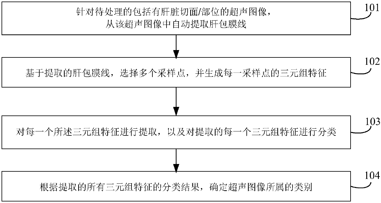
[0103] Such as figure 1 as shown, figure 1 It shows a schematic flowchart of a feature classification method for liver ultrasound images provided by an embodiment, and the method of this embodiment includes the following steps:

[0104] 101. For an ultrasound image including liver sections / parts to be processed, automatically extract the liver capsule line from the ultrasound image.

[0105] For example, step 101 may include:

[0106] The first step, for the ultrasound image to be processed including liver sections / parts, use a sliding window detector to process the ultrasound image, and establish multiple channels in the image block corresponding to the window of the sliding window detector, from the established Extracting pre-selected random rectangular features from multiple channels to obtain a detection response map; the random rectangular features are determined in advance through training samples;

[0107] In the second step, a complete liver envelope line is extract...

Embodiment 2

[0199] The method of the present embodiment includes steps not shown in the following figures:

[0200] Step 601. Obtain the liver capsule line in the ultrasound images of the liver marked as lesions and normal training samples;

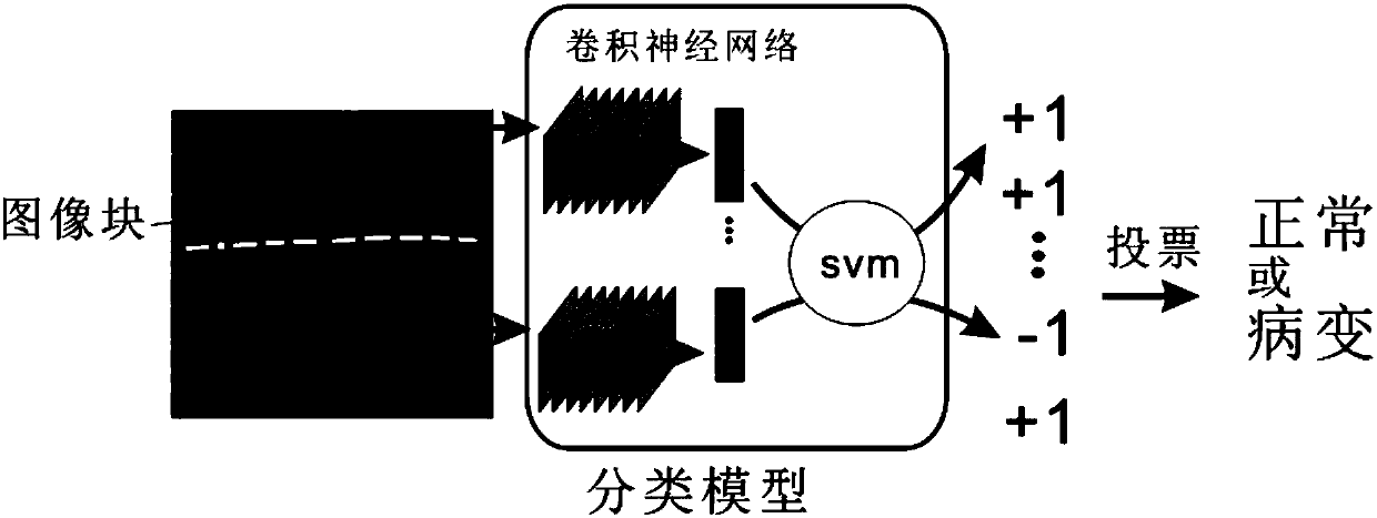
[0201] Step 602, select a certain number of sampling points on each liver envelope line in step 601, three adjacent sampling points form a group, intercept the image block, extract features, and train the support vector machine SVM classifier;

[0202] Step 603, randomly select a certain number of sampling points in the area above the liver envelope line in the training sample in step 601, select three image blocks of different sizes at each sampling point, extract features, and train the SVM classification device;

[0203] Step 604, randomly select a certain number of sampling points in the area below the liver capsule line in the training sample in step 601, select three image blocks of different sizes at each sampling point, extract features, and...


PUM


Abstract

The invention provides a feature classification method for a liver ultrasonic image. The method comprises the steps of S1, for a to-be-processed ultrasonic image comprising a liver section/part, automatically extracting a liver capsule line from the ultrasonic image; S2, based on the extracted liver capsule line, selecting multiple sampling points, and generating a triple feature of each samplingpoint; S3, extracting each triple feature, and classifying the extracted triple features; and S4, according to a classification result of all the extracted triple features, determining the type of theultrasonic image. According to the method, classification results of all image blocks are combined to obtain an accurate classification result; the noise interference is reduced; automatic identification and classification are realized; and the labor cost is reduced.

Description

technical field [0001] The invention relates to image image analysis technology, in particular to a method for feature classification of liver ultrasound images. Background technique [0002] The liver is an organ in the body that mainly has metabolic functions, and it plays the roles of deoxidation, storage of glycogen, and synthesis of secreted proteins in the body. [0003] Liver cirrhosis is a common clinical chronic progressive liver disease, which is caused by diffuse liver damage caused by one or more pathogenic factors such as viral hepatitis, chronic alcoholism, malnutrition, and intestinal infection for a long time or repeatedly , can be complicated by splenomegaly, ascites, edema, jaundice, esophageal varices, hemorrhage, hepatic coma, can develop into liver cancer, and has a high mortality rate. [0004] Timely detection and treatment with drugs can delay the progress of liver cirrhosis, reduce the incidence of liver cancer, improve the long-term survival rate, ...

Claims


Application Information

Patent Timeline
Patent Type & Authority Applications(China)
IPC IPC(8): G06K9/62G06K9/46G06T7/00
CPCG06T7/0012G06T2207/30056G06T2207/20081G06T2207/20084G06V10/462G06F18/2411G06F18/214
Inventor 刘翔
Owner 北京华想联合科技有限公司
Why Eureka
Try Eureka