Single-reagent heparin binding protein detection reagent kit and method for preparing same
A technology of heparin-binding protein and detection kit, which is applied in measurement device, scattering characteristic measurement, color/spectral characteristic measurement, etc., can solve the problems of detection reagent sensitivity, specificity, and unsatisfactory linear range, and achieve a wide measurement linear range. , Simple operation, the effect of simplifying the test program
- Summary
- Abstract
- Description
- Claims
- Application Information
AI Technical Summary
Problems solved by technology
Method used
Image

Examples
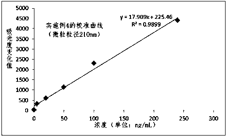
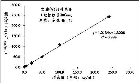
Embodiment 1
[0045] 1) Preparation of latex reagent (300nm, reaction solution) labeled with combination of monoclonal antibody and polyclonal antibody
[0046] Polystyrene latex particles with a carboxyl group on the surface (classification number K030, manufacturer Merck Millipore) with a particle size of 300 nm were labeled by the intermediate ester method. Dilute the microparticles with a particle size of 300nm and carboxyl groups on the surface to 1% in MES buffer solution, and stir at room temperature. Add EDC and Sulfo-NHS solid powder, stir for 2 hours and then centrifuge at 10000RPM for 20 minutes. The latex was washed twice with MES buffer solution, resuspended and divided into two parts. One part was added with HBP monoclonal antibody, stirred and reacted for 2 hours, and the other part was added with HBP polyclonal antibody, stirred and reacted for 2 hours. Both latexes were then centrifuged at 10,000 RPM for 20 minutes, and the supernatant was discarded. The two sets of prec...
Embodiment 2
[0054] 1) Preparation of a pair of monoclonal antibody-labeled latex reagents (200nm, reaction solution)
[0055]200nm polystyrene latex particles with carboxyl groups on the surface were labeled by the intermediate ester method (classification number H20021C, manufacturer Holmes). Microparticles with a particle size of 200 nm were diluted to 1% in PBS buffer solution and stirred at room temperature. Add EDC and Sulfo-NHS solid powder, stir for 2 hours and then centrifuge at 10000RPM for 20 minutes. A pair of HBP monoclonal antibodies was added, and the reaction was stirred for 2 hours. The latex was then centrifuged at 10,000 RPM for 10 minutes, and the supernatant was discarded. The precipitated latex was resuspended in the stock solution and ultrasonically dispersed, stirred for 1 hour before use. In this example, the reaction solution includes latex particles, buffer solution, salt, stabilizer, suspending agent, preservative and surfactant, wherein the concentration of ...
Embodiment 3
[0059] 1) Preparation of polyclonal antibody-labeled latex reagent (120nm, reaction solution)
[0060] 120nm polystyrene latex particles (classification number PS19021, manufacturer Merck Millipore) were labeled by physical adsorption. Microparticles with a particle size of 120 nm were diluted to 1% in PBS buffer solution and stirred at room temperature. Add EDC and Sulfo-NHS solid powder, stir for 2 hours and then centrifuge at 10000RPM for 20 minutes. Add HBP polyclonal antibody, and stir for 2 hours. The latex was then centrifuged at 10,000 RPM for 10 minutes, and the supernatant was discarded. The precipitated latex was resuspended in the stock solution and ultrasonically dispersed, stirred for 1 hour before use. In this example, the reaction solution includes latex particles, buffer solution, salt, stabilizer, suspending agent and preservative, wherein the concentration of latex particles is 0.05%, the buffer solution is 100mM Hepes pH=7.8, and the stabilizer is 1% bov...
PUM
| Property | Measurement | Unit |
|---|---|---|
| Diameter | aaaaa | aaaaa |
| Particle size | aaaaa | aaaaa |
| Particle size | aaaaa | aaaaa |
Abstract
Description
Claims
Application Information