
A microfluidic chip for cell tissue culture and real-time monitoring and its application method

A microfluidic chip, cell tissue technology, applied in tissue cell/virus culture devices, methods of stress-stimulated microbial growth, fluid controllers, etc., can solve the problem of limited development and application, lack of statistical significance, difficulty in high-pass Quantitative experiments and other issues to achieve high practical value

Active Publication Date: 2021-06-25
SOUTHEAST UNIV
View PDF6 Cites 0 Cited by
  • Summary
  • Abstract
  • Description
  • Claims
  • Application Information

AI Technical Summary

Problems solved by technology

[0006] The development and application of microfluidic tissue chips currently used for tissue culture or drug testing are limited in the following aspects: the lack of effective means to precisely manipulate cells and grow them into three-dimensional spherical tissues; the current real-time Detection is generally achieved through microscopic imaging, and phototoxicity has a certain impact on the physiological state of cells and tissues; it is difficult to perform high-throughput experiments, and the sample data is small, lacking statistical significance; tissue samples are not easy to take out for subsequent research and experiments

Method used


Image

  • A microfluidic chip for cell tissue culture and real-time monitoring and its application method
  • A microfluidic chip for cell tissue culture and real-time monitoring and its application method
  • A microfluidic chip for cell tissue culture and real-time monitoring and its application method

Examples


Embodiment Construction

[0041] The technical scheme illustrating the present invention will be described in detail below through specific examples and accompanying drawings. Those skilled in the art can easily understand other advantages and effects of the present invention from the contents disclosed in this specification. The present invention can also be implemented or applied through other different specific implementation modes, and various modifications or changes can be made to the details in this specification based on different viewpoints and applications without departing from the principle of the present invention.

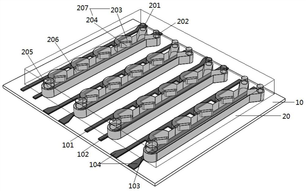
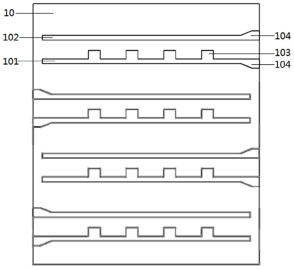
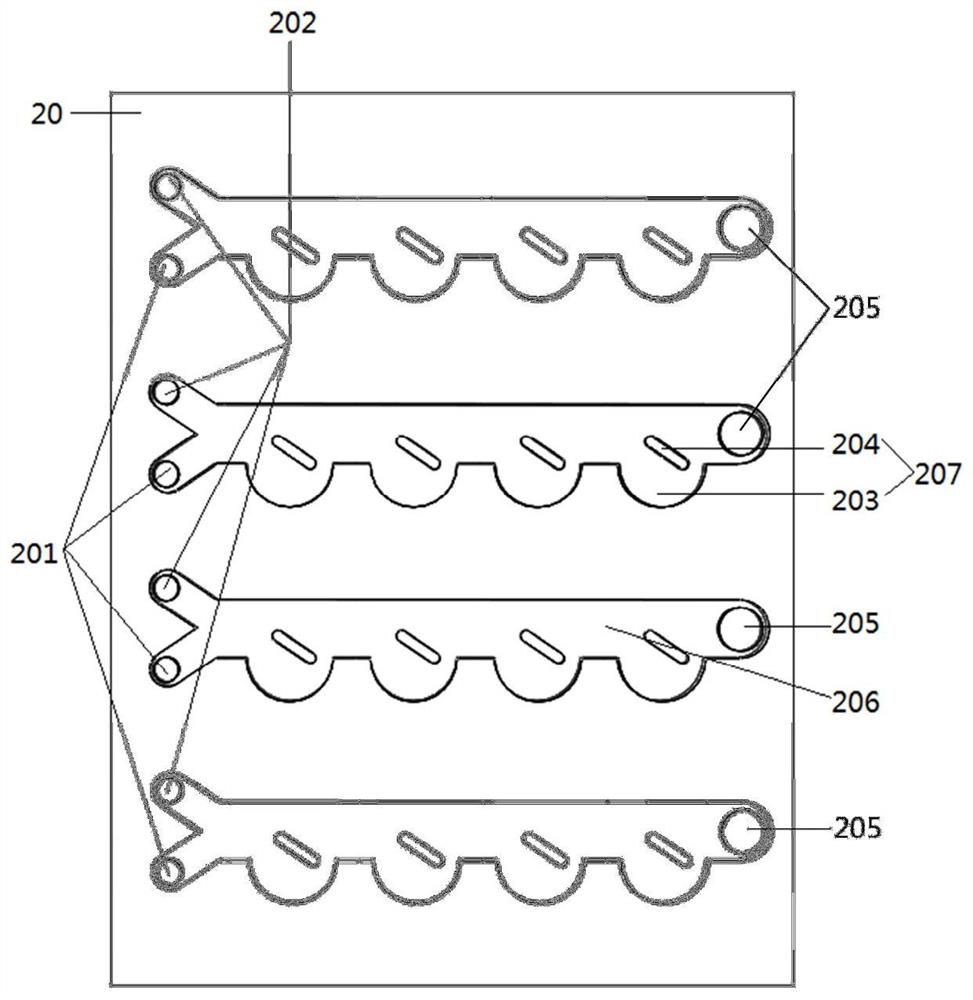
[0042] see Figure 1 to Figure 7 , It should be noted that the diagrams provided in this embodiment are only used to illustrate the basic principles, component structures, working processes and effects of the present invention, so only the components related to the present invention are shown in the diagrams instead of according to the The number, formation and dimensional dr...


PUM


Abstract

The invention discloses a microfluidic chip for cell tissue culture and real-time monitoring and a method for using the chip. The chip includes a glass substrate layer and a PDMS microfluidic channel layer on the glass substrate layer. The glass substrate layer includes a glass substrate and a glass substrate. Several pairs of microelectrodes arranged on the glass substrate layer, the PDMS microfluidic channel layer includes several independent microfluidic channels, and the microelectrode pairs on the glass substrate correspond to the microfluidic channels on the PDMS microfluidic channel layer; The microelectrodes are electrically connected to external circuits. The methods of use include cell capture, cell tissue culture, electrical impedance spectroscopy detection, and tissue release. The chip of the present invention is processed with a transparent base material, and the microscopic imaging can be used as a supplementary means for electrical impedance spectroscopy detection to observe and analyze biological processes such as dynamic growth and physiological behavior of cell tissues in situ. It can be used in fields such as biological research of cells or tissues and drug screening.

Description

technical field [0001] The invention relates to microfluidic technology and a biological cell tissue culture device, in particular to a microfluidic chip for cell tissue culture and real-time monitoring and its application method. Background technique [0002] Before the official application of new drugs, animal experiments and human clinical trials are generally required. Due to the huge difference between animals and humans, the information provided in animal experiments is unreliable, and animal experiments involve ethical issues, while human clinical trials will bring potential harm to the bodies of experimental volunteers due to unstable factors such as side effects of drugs. On the other hand, there are huge uncertainties and risks of failure. Therefore, the dynamic culture of in vitro biological tissues and drug experiments are important means to solve the problems of long drug development cycle and high cost. [0003] The in vitro culture of cells is an important t...

Claims


Application Information

Patent Timeline
Patent Type & Authority Patents(China)
IPC IPC(8): C12M3/02C12M1/42C12M1/34
CPCC12M21/08C12M23/16C12M23/22C12M27/20C12M41/36C12M41/46B01L3/502715B01L3/502761B01L2400/0424B01L2400/0457B01L2400/086C12M45/07B01L3/50273B01L2300/0645B01L2300/0864B01L2400/0475
Inventor 朱真厉俏单金鼎鑫俞峰耿杨烨
Owner SOUTHEAST UNIV
Learn More