Detecting method for circulating tumor cell surface marker molecule PD-L1

A PD-L1 and tumor cell technology, applied in the field of molecular biology, can solve the problems of patient injury, PD-L1 confusion, inconsistent staining technology and conditions, and achieve the effect of simple detection method, high sensitivity and good specificity

Inactive Publication Date: 2018-09-07
THE FIRST AFFILIATED HOSPITAL OF SOOCHOW UNIV
View PDF5 Cites 13 Cited by
  • Summary
  • Abstract
  • Description
  • Claims
  • Application Information

AI Technical Summary

Problems solved by technology

At present, the detection of PD-L1 in my country is relatively chaotic. First, the staining techniques and conditions are not uniform; second, the staining antibodies are diverse; third, the pathological evaluation standards are no

Method used


Image

  • Detecting method for circulating tumor cell surface marker molecule PD-L1

Examples


Embodiment 1

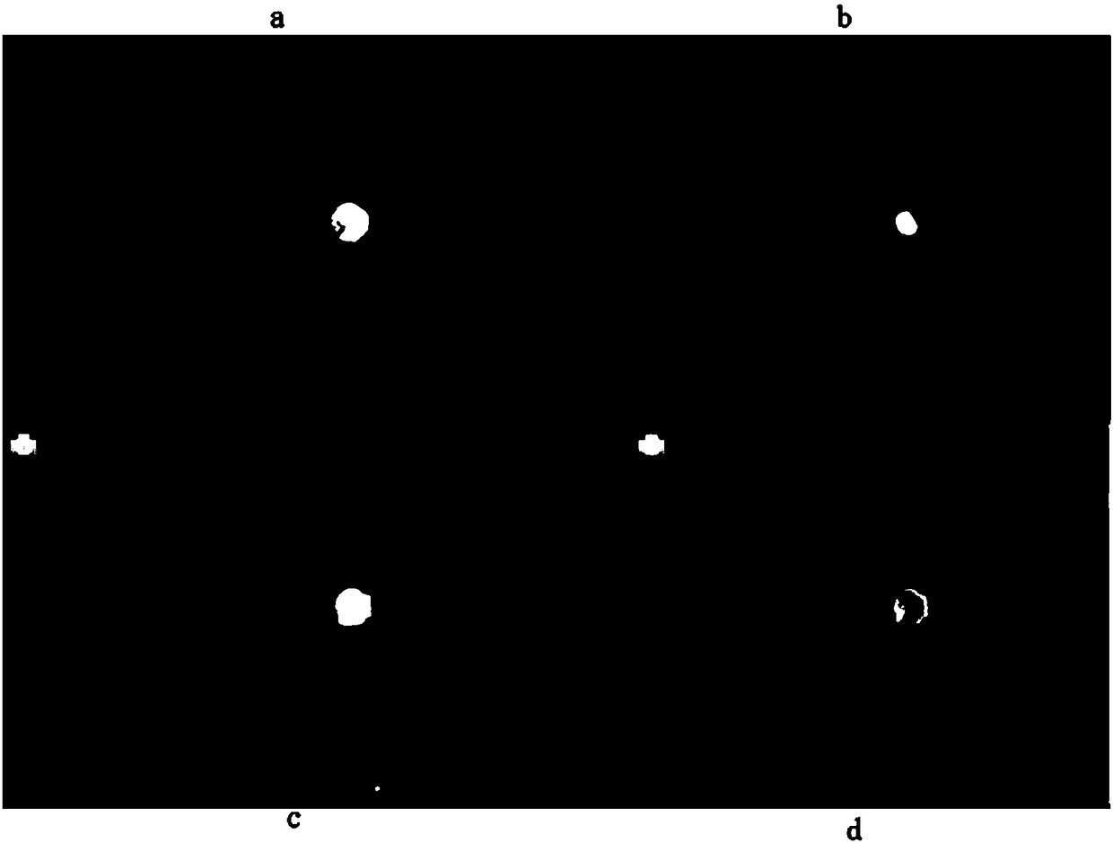
[0022] figure 1 It is a schematic diagram for detecting the positive expression of PD-L1 in circulating tumor cells. figure 1 a is the blue fluorescent channel, use DAPI to mark the nucleus, if the blue color is DAPI+, it means it is a complete cell, if it does not show blue color, it is DAPI-, it is not a complete cell; figure 1 b is the green fluorescent channel, marked with PD-L1 protein, if it is green, it is PD-L1+, and if it is not green, it is PD-L1-; figure 1 c is the red fluorescent channel, marking CK protein, CK+ when it is red, and CK- when it is not. figure 1 d is the synthesis of the three channels.

[0023] This example is an example of detecting the expression of PD-L1 on the surface of circulating tumor cells in clinical liver cancer samples, which specifically includes the following steps:

[0024] 1) Whole blood processing:

[0025] a. Add 200 μL of 1× red blood cell lysate to 2 mL of whole blood, place at room temperature for 15 min, and shake evenly du...


PUM


Abstract

The invention discloses a detecting method for a circulating tumor cell surface marker molecule PD-L1. The detecting method comprises the following steps: (1) treating whole blood with red blood celllysate to separate out karyocyte, and fixing with formaldehyde; (2) performing positive screening through tumor immune fluorescent marker cytokeratin antibody anti-CK, performing PD-L1 primary antibody incubation on all cells, and performing PD-L1 secondary antibody incubation with a fluorescence group marked with FITC, and marking all cells through cell nucleus fluorescent dye DAPI; and (3) adopting high-flux multi-color imaging analysis to select light filter plates of CY5, FITC and DAPI, observing fluorescent color on the surface of a channel, and finally realizing detection on the circulating tumor cell surface marker molecule PD-L1. The detecting method is simple and convenient, is quick, is economical, is non-invasive, is high in sensitivity and is good in specificity.

Description

technical field [0001] The invention belongs to the technical field of molecular biology, in particular to a method for detecting the surface marker molecule PD-L1 of circulating tumor cells. Background technique [0002] Programmed death receptor-1 (PD-1) is the main immune checkpoint receptor, by binding to its ligand programmed death ligand-1 (PD-L1), it down-regulates the effector function of T cells, thus contributing to To maintain tolerance to tumor cells. Blocking these pathways by anti-PD-1 and anti-PD-L1 antibodies may help prevent this downregulation and allow T cells to maintain their antitumor properties and ability to mediate tumor cell death. There are currently three main inhibitors of PD-1 and PD-L1 on the market, pembrolizumab (trade name: Keytruda), Nibolumab (trade name: Opdivo) and Atezolizumab (trade name: Tecentriq), which can be used for melanoma, non-small cell Treatment of various cancers such as lung cancer and bladder cancer. However, not all p...

Claims


Application Information

Patent Timeline
IPC IPC(8): G01N21/64
CPCG01N21/6428G01N21/6486
Inventor 马海涛黄海涛陈少慕张标顾媛媛雷豪志王煜蘅祝琳
Owner THE FIRST AFFILIATED HOSPITAL OF SOOCHOW UNIV
Why Eureka
Try Eureka