Methods for preparing cutting guide plates and determining cut soft tissue substituting flap volume by using digital space reconstruction and 3D printing technology

A 3D printing and spatial reconstruction technology, which is applied in the field of medical treatment, can solve the problems of high personal requirements, insufficient space, surface skin collapse, etc.

Active Publication Date: 2018-10-19
ZHEJIANG CANCER HOSPITAL
View PDF10 Cites 2 Cited by
  • Summary
  • Abstract
  • Description
  • Claims
  • Application Information

AI Technical Summary

Problems solved by technology

Therefore, how to accurately determine the size of the cutting guide plate has become a technical problem (in the prior art, the cutting guide plate is generally not established by building a three-dimensional model, but the doctor’s personal experience is used to prepare the cutting guide plate and perform skin flap removal. Surgeons have high personal requirements, which is a challenge for inexperienced novices)
[0007] On the other hand, in the prior art, the repair of soft tissue defects generally only focuses on the repair of the surface layer, but does not focus on the repair of subcutaneous soft tissue vacancies.
If the resection surface...

Method used


Image

  • Methods for preparing cutting guide plates and determining cut soft tissue substituting flap volume by using digital space reconstruction and 3D printing technology
  • Methods for preparing cutting guide plates and determining cut soft tissue substituting flap volume by using digital space reconstruction and 3D printing technology
  • Methods for preparing cutting guide plates and determining cut soft tissue substituting flap volume by using digital space reconstruction and 3D printing technology

Examples


Embodiment 1

[0064] Patient Lu **, male, 34 years old.

[0065] The right tongue ulcer appeared more than 2 months ago, and she went to the local hospital for treatment, considering tongue ulcer and anti-inflammatory treatment. In the past one month, the mass has increased significantly, and the pain has increased.

[0066] Biopsy: "Tongue" consistent with well-differentiated squamous cell carcinoma.

[0067] Examination: The right tongue mass was about 4cm in size, ulcerated, with limited tongue movement, and multiple enlarged lymph nodes in the right upper neck.

[0068] Diagnosis: Malignant tumor of the tongue

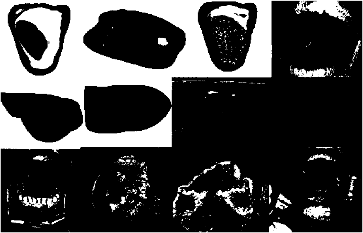
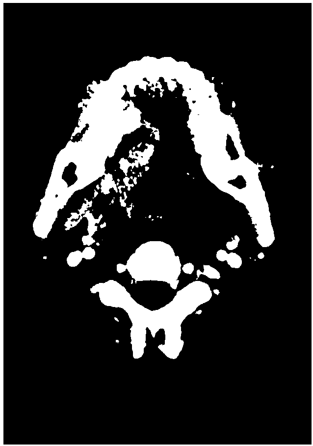
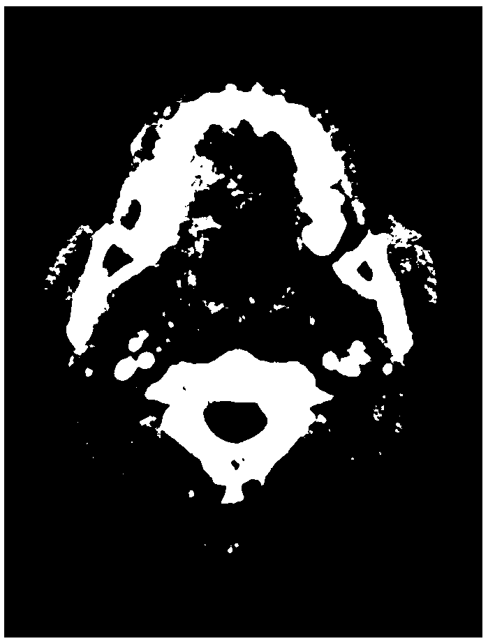
[0069] Such as Figure 1-4 As shown, the surgery includes the following steps:

[0070] 1) Obtain the patient's thin-slice CT data (thickness 0.2mm), convert it into DICOM data format and import it into the 6D dental implant design software (6D-DENTAL Tech Co, Ltd, China) to reconstruct the soft tissue model shape and tumor shape of the patient area, Obtain editable .stl fo...

Embodiment 2

[0078] Patient Cai**, male, 58 years old.

[0079] 2016.5 Oral cavity: squamous cell carcinoma of the floor of the mouth (moderately differentiated). Esophagus: Poorly differentiated (squamous) carcinoma of the esophagus 29-31cm from the incisors (esophagus).

[0080] 2016-8-10 "1. Extensive excision of the malignant mass on the floor of the mouth; 2. Partial resection of the malignant mass of the tongue; 3. Neck dissection (bilateral); 4. Tracheotomy; 5. Mandible Partial amputation (titanium plate reconstruction); 6. Right pectoralis major muscle reconstruction; 7. Mouth floor reconstruction".

[0081] Postoperative pathology: 1. (Floor of mouth) high-moderately poorly differentiated squamous cell carcinoma (tumor size 4.5×3.5×2.7cm), infiltrating into submucosal and striated muscle tissue, and invading salivary gland, mandible and nerve tissue. 2. Chronic inflammation of lymph nodes in 10 (left neck II, III) and 23 (right neck), see also some submandibular gland tissues. ...

Embodiment 3

[0096] The difference between this embodiment and Embodiment 1 is that the three-dimensional model is first printed as a curved surface physical model, which specifically includes the following steps:

[0097] 1) Obtain thin-slice (0.2mm) CT data of the patient through CT scanning, then convert it into DICOM data format and import it into the 6D dental implant design software (6D-DENTAL Tech Co, Ltd, China) to reconstruct the soft tissue model shape and Tumor morphology, obtain editable model data in .stl format.

[0098] 2) Combining magics design software (Materialise Ltd, The Kingdom Of Belgium) and Geomagice Studio design software (Geomagice, USA) to edit the 3D model, plan the repair plan, and consider the safe range of tumor resection to demarcate the lesion tissue resection boundary and determine the resection lesion area; then the repaired surface is extracted by the GeomagiceStudio design software, calculated and converted into a Nurbs surface, and saved as data in a ...


PUM


Abstract

The invention relates to the medical science field, and discloses methods for preparing cutting guide plates and determining a cut soft tissue substituting flap volume by using a digital space reconstruction and 3D printing technology. The method includes the following steps: 1) building a three-dimensional model through CT scanning; 2) determining an excision lesion area, and extracting a repaircurved surface; 3) converting the curved surface into a plane; and 4) preparing a cutting guide plate through 3D printing. Substituting flap can be accurately cut in a donor site by using the cuttingguide plate. The method for determining the substituting flap volume includes the following steps: a) building a three-dimensional model through the CT scanning; b) determining an excision lesion area, and calculating the volume of an excision part; c) adding and soaking the substituting flap in a harmless liquid to calculate volume increment; and d) performing trimming on the subcutaneous soft tissue of the substituting flap so that an exact volume can be obtained. The volume of soft tissue defect prosthesis also needs digital assessment. The accurate assessment of the substituting flap volume can make the appearance of the prosthesis plim after surgery.

Description

technical field [0001] The invention relates to the field of medical treatment, in particular to a method for preparing a cutting guide plate and determining the volume of cut soft tissue instead of a skin flap by using digital space reconstruction and 3D printing technology. Background technique [0002] After tumor resection, the space left by the resected soft tissue needs to be filled and repaired, and repairing complex soft tissue defects is extremely challenging for surgeons. Currently, the main methods for repairing soft tissue defects include pedicled flaps, free flaps, and artificial substitutes. At present, it is required to retain more functional peripheral soft tissue under the premise of safe tumor resection margins, and try to restore the shape, function and tissue volume of the reconstructed organs as much as possible. At the same time, there is often a lack of precise preoperative design and intraoperative strain when faced with cases of large-area defects i...

Claims


Application Information

Patent Timeline
IPC IPC(8): A61B17/322
CPCA61B17/322A61B34/10A61B2017/3225A61B2034/102A61B2034/107A61B2034/108B33Y10/00B33Y80/00
Inventor 葛明华徐加杰陈超兰霞斌刘云峰于长洋孙晶晶李升谭卓朱秀
Owner ZHEJIANG CANCER HOSPITAL
Why Eureka
Try Eureka