Lesion identification method and device, computer device, and readable storage medium
A recognition method and part technology, applied in the field of image processing, to achieve the effect of improving accuracy
- Summary
- Abstract
- Description
- Claims
- Application Information
AI Technical Summary
Problems solved by technology
Method used
Image
Examples
Embodiment 1
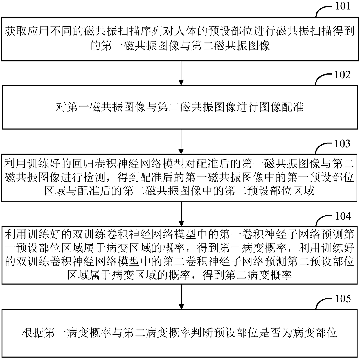
[0062] figure 1 It is a flow chart of the method for identifying lesion parts provided by Embodiment 1 of the present invention. The lesion identification method is applied to a computer device. The lesion identification method performs lesion identification according to different sequences of magnetic resonance images, determines whether the preset site is a lesion, and determines the specific location of the lesion.
[0063] like figure 1 As shown, the lesion identification method specifically includes the following steps:
[0064] Step 101 , acquiring a first magnetic resonance image and a second magnetic resonance image obtained by performing magnetic resonance scanning on preset parts of a human body using different magnetic resonance scanning sequences.
[0065] MRI (Magnetic Resonance Imaging, Magnetic Resonance Imaging) image is one of the commonly used medical images. MRI imaging is a type of tomography. It uses magnetic resonance phenomena to obtain electromagneti...
Embodiment 2
[0124] Figure 4 It is a structural diagram of a lesion identification device provided in Embodiment 2 of the present invention. like image 3 As shown, the lesion identification device 10 may include: an acquisition unit 401 , a registration unit 402 , a detection unit 403 , a prediction unit 404 , and a judgment unit 405 .
[0125] The obtaining unit 401 is configured to obtain a first magnetic resonance image and a second magnetic resonance image obtained by performing magnetic resonance scanning on preset parts of the human body using different magnetic resonance scanning sequences.
[0126] MRI (Magnetic Resonance Imaging, Magnetic Resonance Imaging) image is one of the commonly used medical images. MRI imaging is a type of tomography. It uses magnetic resonance phenomena to obtain electromagnetic signals from the human body and reconstruct human body information to obtain MRI images. .
[0127] In a specific embodiment, the method for identifying a lesion site can be ...
Embodiment 3
[0185] Figure 5 It is a schematic diagram of a computer device provided by Embodiment 3 of the present invention. The computer device 1 includes a memory 20 , a processor 30 and a computer program 40 stored in the memory 20 and executable on the processor 30 , such as a lesion recognition program. When the processor 30 executes the computer program 40, it realizes the steps in the above embodiment of the method for identifying lesion parts, for example figure 1 Steps 101-105 are shown. Alternatively, when the processor 30 executes the computer program 40, it realizes the functions of the modules / units in the above device embodiments, for example Figure 4 Units 401-405 in.
[0186]Exemplarily, the computer program 40 can be divided into one or more modules / units, and the one or more modules / units are stored in the memory 20 and executed by the processor 30 to complete this invention. The one or more modules / units may be a series of computer program instruction segments c...
PUM
 Abstract
Description
Claims
Application Information
 - R&D
- Intellectual Property
- Life Sciences
- Materials
- Tech Scout
- Unparalleled Data Quality
- Higher Quality Content
- 60% Fewer Hallucinations
Browse by: Latest US Patents, China's latest patents, Technical Efficacy Thesaurus, Application Domain, Technology Topic, Popular Technical Reports.
© 2025 PatSnap. All rights reserved.Legal|Privacy policy|Modern Slavery Act Transparency Statement|Sitemap|About US| Contact US: help@patsnap.com


