
Immunochromatographic test strip for quantitatively detecting canine C-reactive protein and preparation method thereof

An immunochromatographic test strip and reactive protein technology, applied in the field of biomedical testing, can solve the problems of insufficient sensitivity, accuracy, and linear range, etc.

Inactive Publication Date: 2018-11-30
海卫特(广州)医疗科技有限公司
View PDF7 Cites 10 Cited by
  • Summary
  • Abstract
  • Description
  • Claims
  • Application Information

AI Technical Summary

Problems solved by technology

Immunofluorescence quantitative rapid detection technology, using double antibody sandwich, the principle and advantages of immunochromatography technology, using fluorescein labeling, so that the detection can be quickly quantified; at the same time, the current fluorescein labeling is conventionally labeled T-line antibody, two concentration points The T / C value gap is completely provided by the difference of the T line signal, so the conventional marking method has certain deficiencies in sensitivity, accuracy, and linear range

Method used


Image

  • Immunochromatographic test strip for quantitatively detecting canine C-reactive protein and preparation method thereof
  • Immunochromatographic test strip for quantitatively detecting canine C-reactive protein and preparation method thereof
  • Immunochromatographic test strip for quantitatively detecting canine C-reactive protein and preparation method thereof

Examples


Embodiment 1

[0050] Embodiment 1, the composition of the immunochromatographic test strip of quantitative detection dog CRP

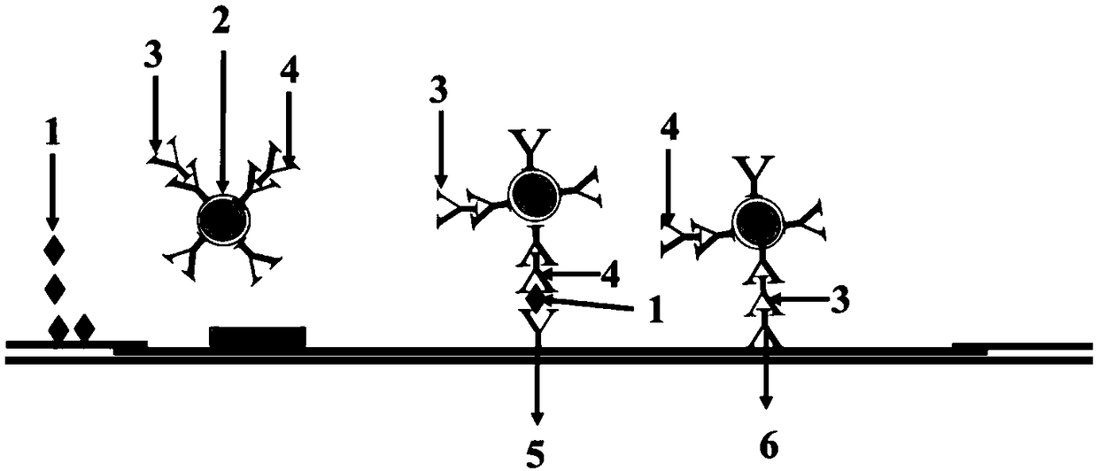
[0051] In the embodiment of the present invention, the canine CRP monoclonal antibody used is a monoclonal antibody prepared by conventional monoclonal antibody technology, and the canine CRP antigen is detected by the double antibody sandwich method. The embodiment of the present invention adopts the mixed labeling technology in which antibodies in the detection area (T line) and the quality control area (C line) are simultaneously labeled on a fluorescent latex particle. The principle is that the T line and the C line compete for the same fluorescent latex particle. When T When the line signal is low, the C line signal is high, and when the T line signal is high, the C line signal is low. This combination mode increases the T / C value of the two concentration points, which can greatly improve the sensitivity and the accuracy of the detection results. Widen the de...

Embodiment 2

[0057] Embodiment 2, the preparation of the immunochromatographic test strip of quantitative detection dog CRP

[0058] In this embodiment, the preparation of the immunochromatographic test strip for quantitatively detecting canine C-reactive protein comprises the following steps:

[0059] 1. Preparation of fluorescent latex particles-canine CRP antibody-goat anti-chicken IgY antibody complex

[0060] 1.1 Activation of fluorescent latex particles

[0061] Ultrasonic treatment of fluorescent latex particles for 30 seconds to adjust the concentration of fluorescent latex particles to 1.0×10 12 / mL, centrifuge at 10,000×g for 5 minutes, dissolve the precipitate with 80 μL of distilled water, and treat with 200W ultrasonic wave for 30 seconds; first add 10 μL of 20 mg / mL carbodiimide, mix well, then add 10 μL of 20 mg / mL N-hydroxysulfur Substitute succinamide, mix well; incubate at room temperature for 20 minutes, centrifuge at 10,000×g for 5 minutes, dissolve the precipitate ...

Embodiment 3

[0072] Embodiment 3, establishment of the reaction mode of the immunochromatographic test strip for quantitatively detecting canine CRP

[0073] 1. Standard curve drawing

[0074] Prepare the canine CRP antigen standard with canine CRP negative serum to prepare a series of concentration standards: 0mg / L, 2.5 mg / L, 5.0mg / L, 10mg / L, 20mg / L, 40mg / L, 80mg / L, 160mg / L , 400 mg / L, take 10 μL dropwise into the CRP sample buffer and mix by inverting repeatedly (8-10 times), then absorb 75 μL of the mixed liquid and dropwise into the sample hole of the test strip, after 3 minutes, pass the quantitative The detection device is Healvet instrument HV-FIA 3000 for detection, read out the signal intensity ratio of the detection area and the quality control area, that is, the T / C value (each concentration point is measured 10 times, and the average value is taken), and the concentration T / C standard curve is drawn .

[0075] 2. ID chip burning

[0076] Draw the corresponding concentratio...


PUM

PropertyMeasurementUnit
diameteraaaaaaaaaa
wavelengthaaaaaaaaaa
concentrationaaaaaaaaaa

Abstract

The invention discloses an immunochromatographic test strip for quantitatively detecting a canine C-reactive protein and a preparation method thereof. The test strip is composed of a sample pad, a coating film and a water absorbent paper, which are sequentially lapped and pasted on a bottom plate. The coating film comprises a labeling zone, a detection zone and a quality control zone. The labelingzone is coated with a canine CRP monoclonal antibody labeled with fluorescent latex particles and a goat anti-chicken IgY antibody and the canine CRP monoclonal antibody and the goat anti-chicken IgYantibody are labeled to the same fluorescent latex particle. The detection zone is coated with another canine CRP monoclonal antibody at an epitope different from the epitope where the canine CRP monoclonal antibody is arranged. The canine CRP monoclonal antibody and the goat anti-chicken IgY antibody are connected to the same fluorescent latex. The detection zone and the quality control zone share the same fluorescent latex particle so that the signal amount of the reaction system is magnified and thus the sensitivity and accuracy are greatly improved and the linear range is broadened.

Description

technical field [0001] The invention belongs to the field of biomedical testing, in particular to an immunochromatographic test strip for quantitatively detecting canine C-reactive protein and a preparation method thereof, which is particularly suitable for the detection of canine CRP. Background technique [0002] C-reactive protein (C-reactive protein, CRP), named after Tillet and Francis found in the serum of patients with acute lobar pneumonia in 1930 that it can react with pneumococcal c polysaccharide in the presence of calcium ions, is an important acute human Phase response protein, the acute phase concentration can be increased by thousands of times, and the half-life of CRP in the circulation is 19 hours. [0003] CRP is a typical representative of acute phase protein. CRP can activate complement and strengthen the phagocytosis of phagocytic cells to play an opsonizing role, thereby removing pathogenic microorganisms invading the body and damaged, necrotic, and ap...

Claims


Application Information

Patent Timeline
Patent Type & Authority Applications(China)
IPC IPC(8): G01N33/68G01N33/577G01N33/569G01N33/558G01N33/533
CPCG01N33/533G01N33/558G01N33/56944G01N33/577G01N33/68G01N2800/52G01N2800/7095
Inventor 何浩王伟
Owner 海卫特(广州)医疗科技有限公司
Learn More