Application of TNFR1 gene and encoded protein thereof
A gene and coding technology, applied in the field of autoimmune disease treatment, can solve the problem of unclear specific roles of inflammatory T cells and regulatory T cells, and achieve the effects of avoiding serious side effects, reducing drug dosage, and promoting differentiation
- Summary
- Abstract
- Description
- Claims
- Application Information
AI Technical Summary
Problems solved by technology
Method used
Image
Examples
Embodiment 1
[0070] Example 1 Extraction of naive CD4 from mouse spleen + T cell
[0071] Naive CD4 was extracted by autoMACS magnetic bead sorting method + T cells. First enrich the spleen cells of the mouse with nylon hair column for T cell enrichment, then add biotin mouse anti-CD8 antibody 6ul, biotin mouse anti-B220 antibody 8ul, biotin mouse anti-CD11b antibody 1.5ul, biotin mouse Anti-CD11c antibody 1.5ul, biotin mouse anti-CD49b 1ul, mix well, and incubate at 4°C for 20 minutes. Then add anti-biotinbeads 8ul, mix well, and incubate at 4 degrees for 15 minutes. The autoMACS instrument performs the first step of negative selection to remove non-CD4 + cell. Add mouse CD62L-beads 8ul and incubate at 4 degrees for 15 minutes. The autoMACS instrument performs the second step of positive selection. The seroselective tube is both naive CD4 + T cells. Detection of naiveCD4 by flow cytometry + T cell purity (>90%).
Embodiment 2
[0072] Example 2 naive CD4 + Induction of T cells into Th1 and Th17 cells in vitro
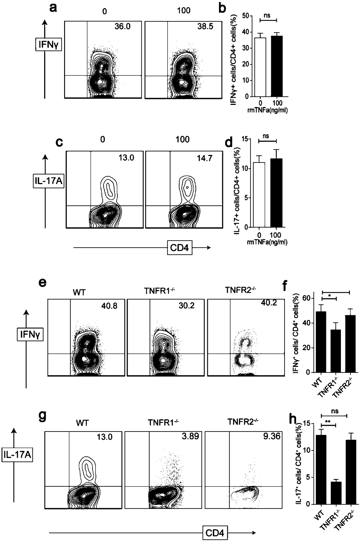
[0073] The extracted WT naive CD4 + T cells were planted in 96-well plates, and CD3 (1ug / ml), CD28 (1ug / ml) and anti-IL-4 monoclonal antibody (5μg / ml) were added to the culture system, with or without rmTNFα. For Th1 cell differentiation, add rmIL-12 (10ng / ml, R&D Systems); for Th17 cells, add rmIL-6 (10ng / ml) and rhTGF-β (2ng / ml), anti-IFN-γ (5μg / ml) ml), the naive CD4 + T cells were cultured in the above system for three days. Collect cells and detect IL-17A using flow cytometry + , IFNγ + expression.
[0074] The result is as figure 1 As shown in a~d, the stimulation of TNFα can not enhance WT naive CD4 +In vitro differentiation of T cells into Th1 and Th17 cells.
[0075] Will be from WT, TNFR1 - / - and TNFR2 - / - Naive CD4 extracted from mouse + T cells were planted in 96-well plate or 48-well plate, and CD3 (lug / ml), CD28 (lug / ml) and anti-IL-4 monoclonal antibody (5 μg / ml) wer...
Embodiment 3
[0076] Example 3 Naive CD4 + Induction of T cells into iTregs in vitro
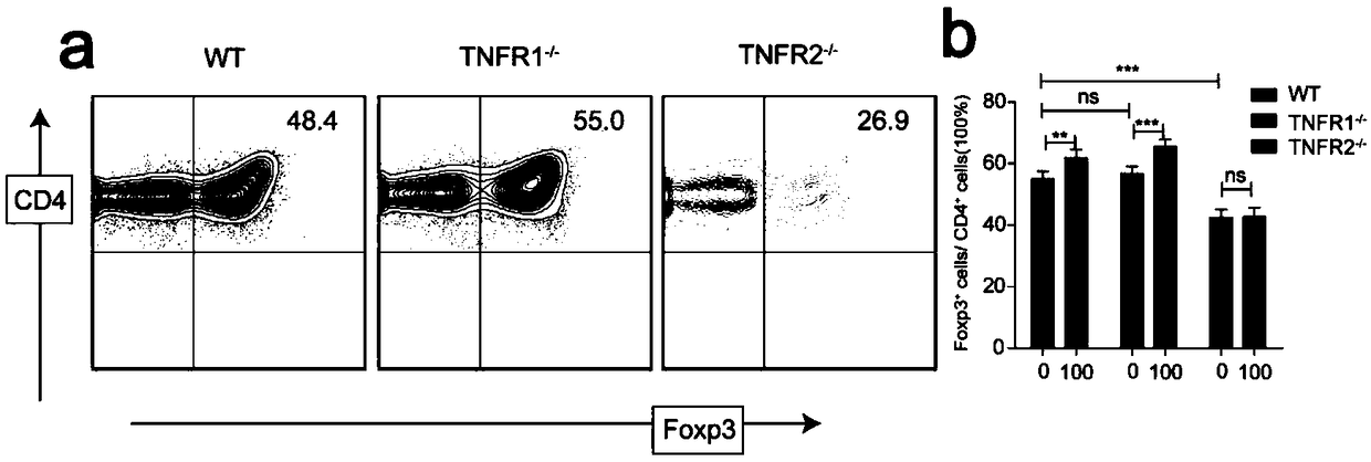
[0077] Will be from WT, TNFR1 - / - and TNFR2 - / - Naive CD4 extracted from mouse + T cells are planted in 96-well plates or 96-well plates, and anti-CD3 / CD28-coated beads (5 cells correspond to one bead), rhIL-2 (50U / ml) and rhTGF-β (2ng / ml) are added to the culture system ), with or without rmTNFα (100ng / ml). Cells were collected at the same time point, and Foxp3 expression was determined by flow cytometry.
[0078] The result is as figure 2 As shown, TNFR1 - / - naive CD4 + T cells can successfully differentiate into iTregs, and when the TNFR2 gene is lost, naive CD4 + The proportion of T cells differentiated into iTregs decreased significantly. Furthermore, rmTNFα fails to promote WT and TNFR1 - / - naive CD4 + Differentiation of T cells to iTregs, response to TNFR2 - / - Cells are useless.
PUM
 Abstract
Description
Claims
Application Information
 - R&D
- Intellectual Property
- Life Sciences
- Materials
- Tech Scout
- Unparalleled Data Quality
- Higher Quality Content
- 60% Fewer Hallucinations
Browse by: Latest US Patents, China's latest patents, Technical Efficacy Thesaurus, Application Domain, Technology Topic, Popular Technical Reports.
© 2025 PatSnap. All rights reserved.Legal|Privacy policy|Modern Slavery Act Transparency Statement|Sitemap|About US| Contact US: help@patsnap.com


