
Fusion method of medical image and image detection method based on fusion medical image learning

A technology of medical imaging and image fusion, applied in the field of deep learning, can solve the problems of not being able to provide pathological tissue information, medical image noise pollution, and affecting medical imaging applications, etc., to enrich comprehensive information, increase training negative samples, and improve confidence Effects of fractional formulas

Active Publication Date: 2022-06-21
HARBIN UNIV OF COMMERCE
View PDF6 Cites 0 Cited by
  • Summary
  • Abstract
  • Description
  • Claims
  • Application Information

AI Technical Summary

Problems solved by technology

[0003] The purpose of the present invention is to provide a medical image fusion method and an image detection method based on fusion medical image learning to solve the problem that medical images are affected by noise pollution, low signal-to-noise ratio, and small grayscale differences between different tissues. application, and technical issues such as single modality images cannot provide richer information on diseased tissue from different angles

Method used


Image

  • Fusion method of medical image and image detection method based on fusion medical image learning
  • Fusion method of medical image and image detection method based on fusion medical image learning
  • Fusion method of medical image and image detection method based on fusion medical image learning

Examples


specific Embodiment approach 1

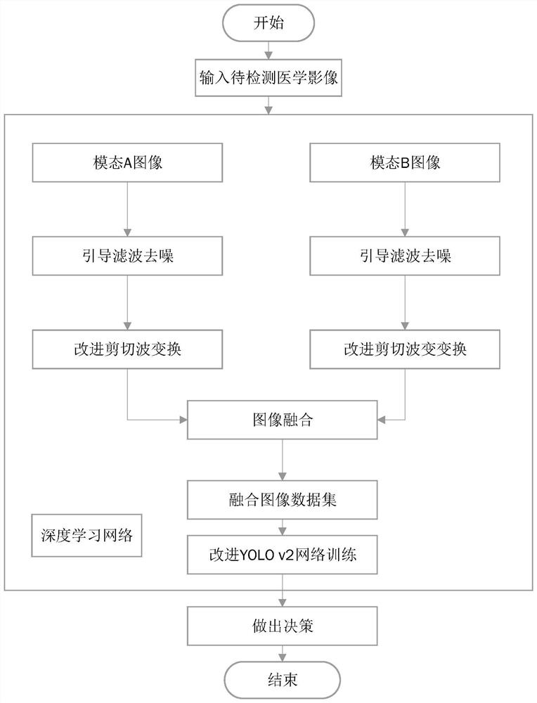
[0061] Specific implementation one: as figure 1 As shown, the fusion method of medical images described in this embodiment includes:

[0062] (1), read mode A medical image I A , Modal B Medical Imaging I B ;

[0063] (2) Preprocess the two types of modal medical images to obtain the denoised image I q ;

[0064] (3) Multi-scale segmentation of the image by using the improved shearlet transform;

[0065] (4) According to the fusion rules, the two types of modal images are fused to obtain the fusion image I F ;

[0066] In step (2), a guided filtering algorithm is used for the preprocessing of the medical image, and the specific process is as follows;

[0067] The input parameters of the guided filtering are the guided image I and the image p (input medical image) that needs to be processed and optimized, and the output is the optimized image q. The guide image and the input image can be preset as I=p, both of which are original medical images. It is derived from the l...

specific Embodiment approach 2

[0100] Specific implementation two: as figure 1 As shown, the image detection method based on fusion medical image learning described in this embodiment includes:

[0101] (1), read mode A medical image I A , Modal B Medical Imaging I B ;

[0102] (2) Preprocess the two types of modal medical images to obtain the denoised image I q ;

[0103] (3) Multi-scale segmentation of the image by using the improved shearlet transform;

[0104] (4) According to the fusion rules, the two types of modal images are fused to obtain the fusion image I F ;

[0105] (5), compose all fused images into a fused image dataset S{I F};

[0106] (6), using the improved YOLO v2 deep learning algorithm to train the data set to generate a training network;

[0107] (7), use the trained network to detect the image to be detected, and give a judgment decision.

[0108] In step (6), the YOLO v2 deep learning algorithm is used to train the fused image data set obtained in step (5), and the followin...


PUM


Abstract

A fusion method of medical images and an image detection method based on fusion medical image learning relate to an image detection technology based on fusion medical image learning. The present invention solves the problem that medical images are polluted by noise, the signal-to-noise ratio is low, and the difference in gray levels between different tissues is small, which affects the application of medical images, and single-mode images cannot provide more information about diseased tissues from different angles. question. Read the two types of modal images and preprocess the two types of modal images respectively to obtain denoising images; use the improved shearlet transform to segment the images in multiple scales; according to the fusion rules, the two types of modal images are processed Fusion to obtain a fusion image; all the fusion images are combined into a fusion image data set; the data set is trained using the improved YOLO v2 deep learning algorithm to generate a training network; the trained network is used for detection. The fusion of medical images of different modalities provides richer information on diseased tissue from different angles.

Description

technical field [0001] The invention belongs to the field of deep learning, and specifically relates to an image detection technology based on fusion medical image learning. Background technique [0002] More than 90% of medical data come from medical images, including ultrasound, computed tomography (CT), nuclear magnetic resonance (MRI), positron emission tomography (PET), etc. Advanced medical image detection technology is used in various diseases, especially tumors. It plays an important role in the detection and diagnosis of diseases. However, as a screening procedure for early diagnosis of tumors, the current imaging diagnosis mainly relies on manual completion. One or more experienced doctors are required to check whether there are signs of lesions and make a diagnosis. In the era of medical big data, the increasing number of images Data brings great difficulties to manual reading, which is not only expensive and time-consuming, but also because of the heavy workload...

Claims


Application Information

Patent Timeline
Patent Type & Authority Patents(China)
IPC IPC(8): G06T5/00G06T5/50G06T7/00
CPCG06T5/50G06T7/0012G06T2207/30096G06T2207/20084G06T2207/20081G06T5/70
Inventor 李鹏张衍儒白世贞任宗伟
Owner HARBIN UNIV OF COMMERCE
Learn More