

Photoacoustic endoscope for high-precision three-dimensional scanning of front micro motor and imaging method
A micro-motor and three-dimensional scanning technology, applied in endoscopy, medical science, sensors, etc., can solve problems that are not suitable for endoscopic imaging, and achieve the effects of small overall size, reduced noise, and high precision
- Summary
- Abstract
- Description
- Claims
- Application Information
AI Technical Summary
Problems solved by technology
Method used
Image

Examples
Embodiment
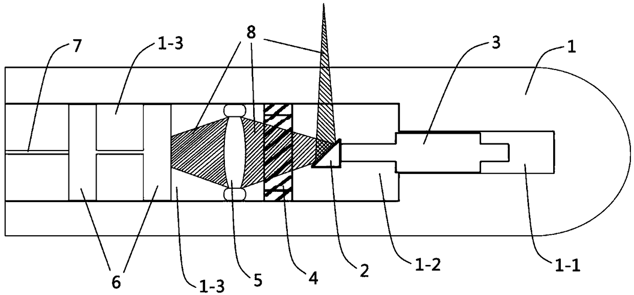
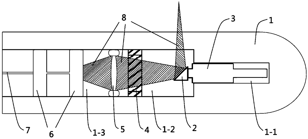
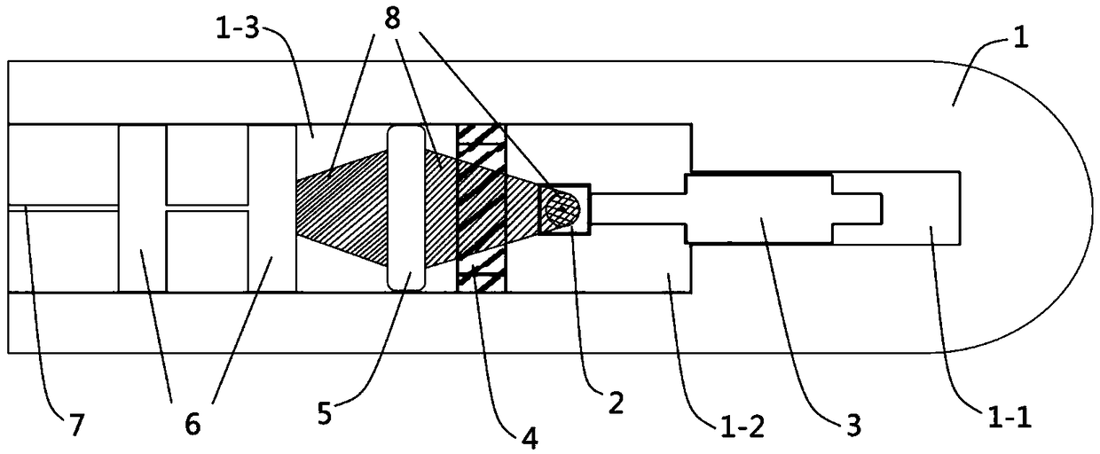
[0035] Such as figure 1 , figure 2 and image 3As shown, a photoacoustic endoscope with high-precision pre-three-dimensional scanning includes a probe shell 1, a micro-motor placement cavity 1-1, an oil chamber 1-2, a cavity 1-3, an optical reflection prism 2, and a micro-motor 3. Ultrasonic transducer 4. Optical zoom lens 5. Optical fiber bearing 6. Optical fiber 7. Laser beam 8.
[0036] The inside of the probe shell 1 is provided with a micro-motor placement chamber 1-1, an oil chamber 1-2 and a cavity 1-3, the micro-motor placement chamber is arranged at the front end of the probe case, and the oil chamber is arranged at the probe The middle position inside the shell, the cavity is arranged at the tail end inside the probe shell.
[0037] The ultrasonic transducer 4 is installed in the middle of the probe shell 1 to separate the oil chamber 1-2 from the cavity 1-3; The main part of the motor 3 is fixed on the micro-motor placement chamber 1-1 at the top of the probe s...
PUM
 Abstract
Description
Claims
Application Information