A kit for predicting the risk of pancreatic cancer postoperative recurrence

A technique for pancreatic cancer and a kit is applied in the field of kits for predicting the risk of postoperative recurrence of pancreatic cancer, which can solve the problems of poor sensitivity and specificity, and achieve the effects of convenient use, high repeatability and strong stability.

Inactive Publication Date: 2019-01-01
ZHONGSHAN HOSPITAL FUDAN UNIV
View PDF5 Cites 0 Cited by
  • Summary
  • Abstract
  • Description
  • Claims
  • Application Information

AI Technical Summary

Problems solved by technology

However, the sensitivity and specificity of the prediction are not good. If a better indicator for predicting the recurrence of pancreatic cancer can be found, it will be of great significance to the treatment and prognosis of pancreatic cancer, and can bring survival benefits to patients.

Method used


Image

  • A kit for predicting the risk of pancreatic cancer postoperative recurrence
  • A kit for predicting the risk of pancreatic cancer postoperative recurrence
  • A kit for predicting the risk of pancreatic cancer postoperative recurrence

Examples


Embodiment 1

[0039] Example 1 Kit for Predicting Postoperative Recurrence Risk of Pancreatic Cancer

[0040] This embodiment provides a kit for predicting the postoperative recurrence risk of pancreatic cancer, including:

[0041] 1) Solution: 4% paraformaldehyde, 0.01M PBS containing 30% sucrose, 0.01M PBS containing 10% donkey serum and 0.2% Triton X-100 in 0.01M PBS, containing 5% donkey serum and 0.2% Triton X -0.01 MPBS of 100;

[0042] 2) Antibody: CD34 antibody (abcam company, #8536), GLUT-1 antibody (abcam company, #18356), immunofluorescence secondary antibody (Invitrogen company, Alexa-555 labeled donkey anti-mouse secondary antibody and Alex-488 labeled donkey anti-rabbit secondary antibody);

[0043] 3) serum: donkey serum (sigma company)

[0044] 4) Water-soluble anti-catabolism mounting agent: Fluoromount-G fluorescent mounting agent (southern biotechnology company);

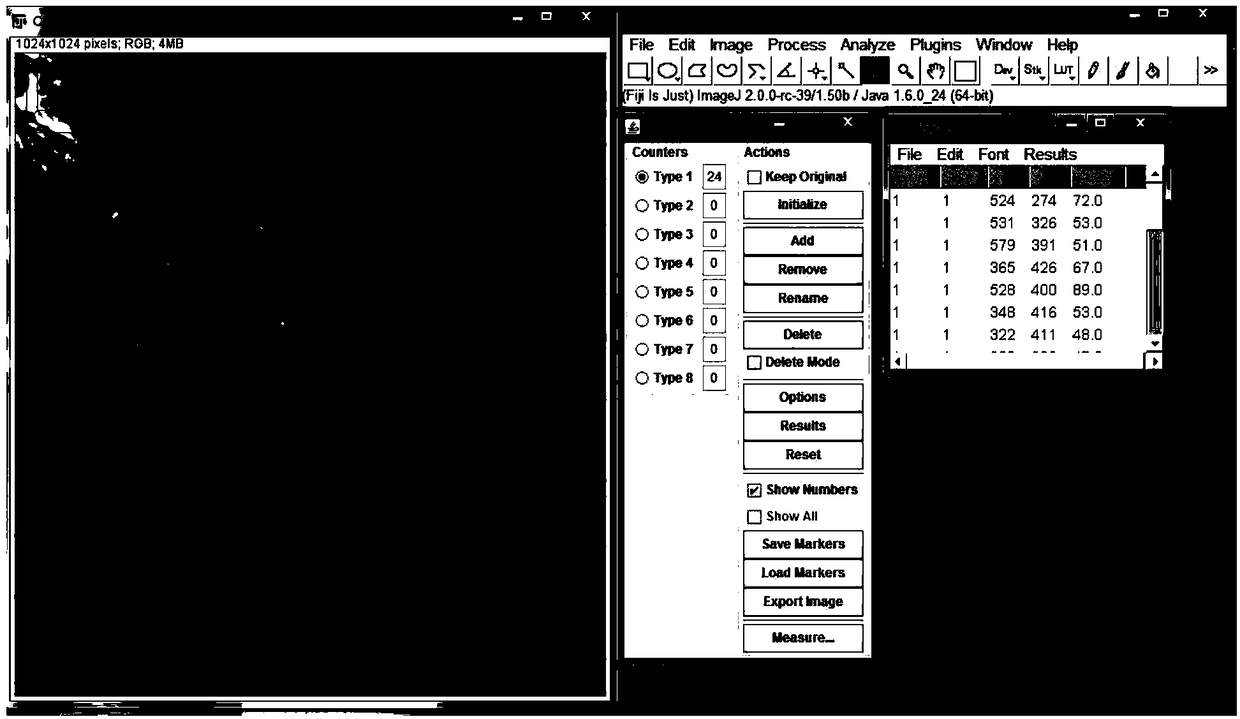
[0045] 5) Image analysis software: Image J;

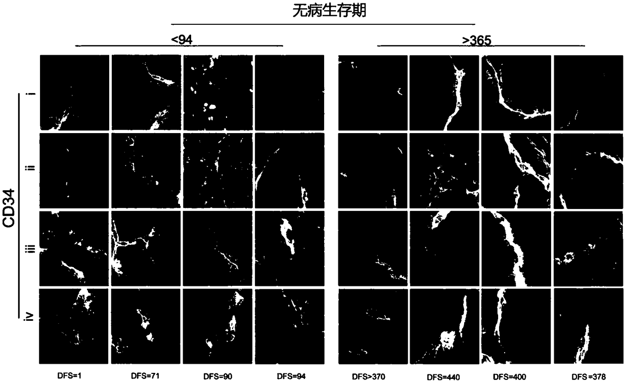
[0046] 6) The average and median length high-risk thresho...

Embodiment 2

[0051] Example 2 Using the kit in Example 1 to process tumor tissue samples

[0052] 1. Collection of human pancreatic cancer tissue samples

[0053] Pathologists or surgeons collect tumor specimens. The length of the collected tumor tissue is 0.5-1.2cm, the width is 0.5-1.2cm, and the thickness is 0.2-0.5cm. When sampling the tumor, it should be as close to the center of the tumor as possible, and the surrounding tissue may not be taken. After sampling, quickly fix in freshly prepared 4% paraformaldehyde (PFA), then store and transport in a 4-degree refrigerator, fix for 24-48 hours, and then put in 30% sucrose / PBS (phosphate buffer solution) Dehydrate overnight.

[0054] 2. Preparation of Thick Sections of Tumor Tissue

[0055] The tumor tissues were taken out from the 30% sucrose / PBS solution, and slices with a thickness of 45 μm were made with a cryostat.

[0056] 3. Section staining

[0057] ①Wash the tissue sections in 0.01mol PBS three times, then place the suspend...

Embodiment 3

[0063] Embodiment 3 image processing and standard curve establishment

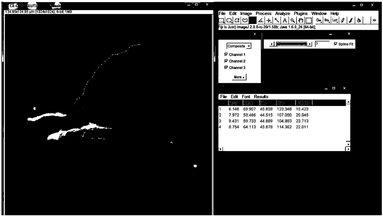
[0064] We used 6-12 CD34 and sugar transporter-1 (GLUT-1) or cytokeratin (cytokeratin, CK) antibody immunofluorescent double-stained sections to scan the basal microvilli of microcirculatory endothelial cells in a patient with pancreatic cancer, and used GLUT- 1 antibody or CK antibody staining to determine the location and location of tumor cells.

[0065] Use a Zeiss 710 laser confocal microscope to read 5-8 immunofluorescent stained tumor sections of a patient. After reading the fluorescent sections, determine the 5-15 microvessels with the most microvessels in the tumor tissue for counting, measurement and calculation.

[0066] Microscope scanning parameters: pixel (pixel): 1024×1024, linear (liner): 2; Z-stack spacing: 1 μm; scanning speed: 5; digital offset (Digital offset): 100-250; Differences in the staining background, so other parameters can be adjusted according to needs.

[0067] Image analy...


PUM

PropertyMeasurementUnit
lengthaaaaaaaaaa
thicknessaaaaaaaaaa
lengthaaaaaaaaaa

Abstract

A kit for predicting the risk of pancreatic cancer postoperative recurrence is disclosed. The kit includes a pancreatic cancer tissue section preparing reagent, a tissue staining reagent, and a lengthhigh-risk threshold and / or density high-risk threshold of basal microvilli of tumor capillary vascular endothelial cells. Immunofluorescence staining and a laser confocal scanning reconstruction method are utilized to reconstruct the complete structure of the basal microvilli of tumor capillary vascular endothelial cells; physical parameters which are density and length of the basal microvilli are measured by software, and are analyzed with postoperative recurrence time of patients with pancreatic cancer; and the average values and medians of the physical parameters which are the length and density of the basal microvilli are utilized to predict the risk of postoperative recurrence for patients with pancreatic cancer. The basal microvilli of capillary vascular endothelial cells only existin malignant tumor, are a pathological technical index, and belong to the category of gold standards for diagnosis of tumor, and therefore, the index has specificity far higher than specificity of other imaging indexes.

Description

technical field [0001] The invention belongs to the field of clinical tumor function detection and pathological diagnosis, and relates to a kit for predicting postoperative recurrence risk of pancreatic cancer. Background technique [0002] Pancreatic cancer is a tumor with a high lethality rate, and its morbidity and mortality are almost the same. Pancreatic cancer is the fourth leading cause of cancer mortality in the United States and fifth in Europe (Siegel et al., 2016). In recent decades, the incidence and mortality of pancreatic cancer have increased rapidly in my country (Machen et al., 2013). According to the 2017 China Cancer Report, pancreatic cancer has become one of the five major tumors that cause death in large cities. Currently, the main treatment for pancreatic cancer is still surgical resection. Although the surgical techniques of pancreatic cancer have been greatly improved, the long-term survival rate after surgery is still very limited (Vincent et al....

Claims


Application Information

Patent Timeline
Patent Type & Authority Applications(China)
IPC IPC(8): G01N21/64G01N1/28G01N1/30
CPCG01N1/286G01N1/30G01N21/6402G01N2001/2873
Inventor 楼文晖韩序
Owner ZHONGSHAN HOSPITAL FUDAN UNIV
Why Eureka
Try Eureka