
An ophthalmic imaging system with automatic retinal feature detection

A feature detection and imaging system technology, applied in eye testing equipment, medical science, diagnosis, etc., can solve problems such as reducing signal quality, and achieve the effect of clear image, promotion of growth, and comprehensive functions

Active Publication Date: 2019-01-04
QINGDAO MUNICIPAL HOSPITAL
View PDF10 Cites 2 Cited by
  • Summary
  • Abstract
  • Description
  • Claims
  • Application Information

AI Technical Summary

Problems solved by technology

Also, since many OCT systems degrade signal quality further by zero latency, flatter retinas may have better intensity uniformity across the B-scan

Method used


Image

  • An ophthalmic imaging system with automatic retinal feature detection
  • An ophthalmic imaging system with automatic retinal feature detection

Examples


Embodiment Construction

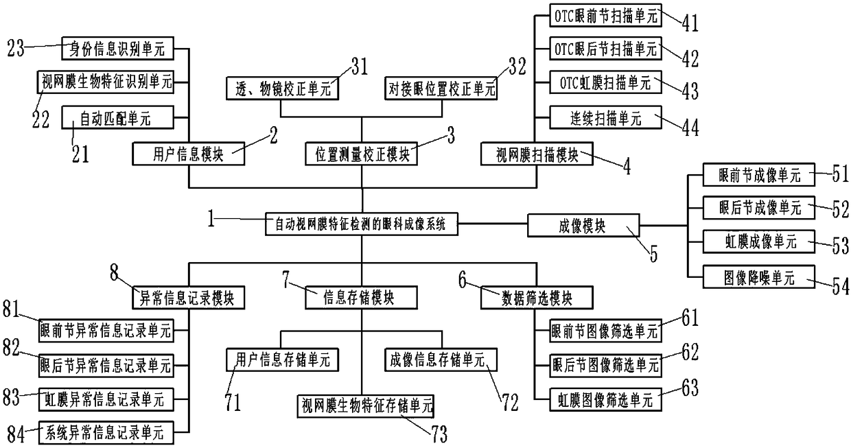
[0026] Attached below figure 1 The present invention is described in further detail with specific examples.

[0027] Such as figure 1 As shown, an ophthalmic imaging system with automatic retinal feature detection mainly includes an ophthalmic imaging system for automatic retinal feature detection 1, a user information module 2, a position measurement correction module 3, a retinal scanning module 4, an imaging module 5, and a data screening module 6. The ophthalmic imaging system 1 for automatic retinal feature detection is respectively connected with the user information module 2, the position measurement correction module 3, the retinal scanning module 4, the imaging module 5, and the data screening module 6;

[0028] The user information module 2 includes an automatic matching unit 21, a retinal biometric identification unit 22, and an identity information identification unit 23; the identity information identification unit 23 is used to check the user identity according ...


PUM


Abstract

The invention discloses an ophthalmic imaging system with automatic retinal feature detection, which mainly comprises a user information module, a position measurement correction module, a retina scanning module, an imaging module, a data screening module, an information storage module and an abnormal information recording module. The system of the invention transmits patient case information through the Internet, protects patient privacy, and provides a communication platform for medical and nursing personnel of related specialties, thereby promoting the growth of related industries. And thesystem function is comprehensive, it can effectively check the anterior retinal segment, posterior retinal segment and iris of the user, and can collect and record the abnormal information of the user. At the same time, the user's identity can be recognized according to the biometrics on the user's retina.

Description

technical field [0001] The invention relates to the technical field of ophthalmology imaging, in particular to an ophthalmology imaging system with automatic retina feature detection. Background technique [0002] Optical coherence tomography (OCT) is a new optical diagnostic technology with the characteristics of non-invasive and non-contact. Existing ophthalmic optical coherence tomography equipment can only image the posterior segment of the eye, such as the examination of the retina, and it also plays an indispensable role in the diagnosis of fundus diseases, especially for the optic nerve (such as optic neuritis, optic atrophy), macular Diseases (such as idiopathic macular hole, macular epiretinal membrane), retinal detachment, medium plasma, medium infiltration, etc. have a greater role in auxiliary diagnosis. Or it can only image the anterior segment, that is, the biological measurement and eye disease research of the anterior segment structures such as the cornea, c...

Claims


Application Information

Patent Timeline
IPC IPC(8): A61B3/14
CPCA61B3/14
Inventor 赵颖姜文静苑伏香周占宇王晓静
Owner QINGDAO MUNICIPAL HOSPITAL
Learn More