System and method suitable for culturing and real-time monitoring of biological tissue

A real-time monitoring and biological tissue technology, applied in biochemical equipment and methods, biological material sampling methods, tissue cell/virus culture devices, etc., to achieve the effect of solving controllability

Active Publication Date: 2019-02-15
REGENOVO BIOTECH
View PDF4 Cites 2 Cited by
  • Summary
  • Abstract
  • Description
  • Claims
  • Application Information

AI Technical Summary

Problems solved by technology

3D bioprinting provides a highly consistent and highly controllable organoid tissue culture method, but in the existing organ-on-a-chip techn

Method used


Image

  • System and method suitable for culturing and real-time monitoring of biological tissue
  • System and method suitable for culturing and real-time monitoring of biological tissue
  • System and method suitable for culturing and real-time monitoring of biological tissue

Examples


example 1

[0115]Step 1. Take a transfer unit 2100, use a bio-3D printer 1000 to print a bio-3D-printed tissue with hepatocytes in it, and put it into a culture medium (DMEM medium+10% fetal bovine serum+1% penicillin streptomycin ) for 20 days, during which the culture medium was replaced every two days;

[0116] Step 2. Take another transfer unit 2100, print a bio-3D printing tissue with tumor cells in it with a bio-3D printer 1000, and put it into a culture medium (DMEM medium+10% fetal bovine serum+1% green chain Mycin) for 20 days, during which the medium was replaced every two days;

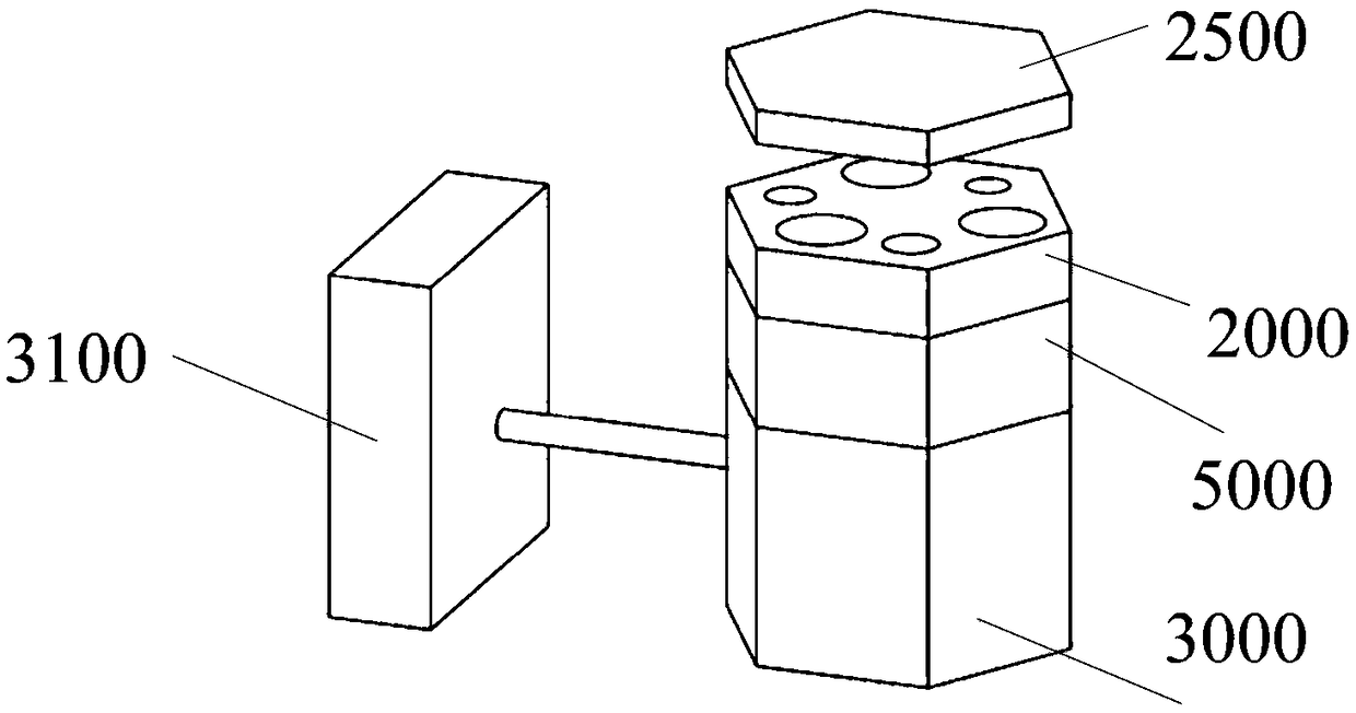
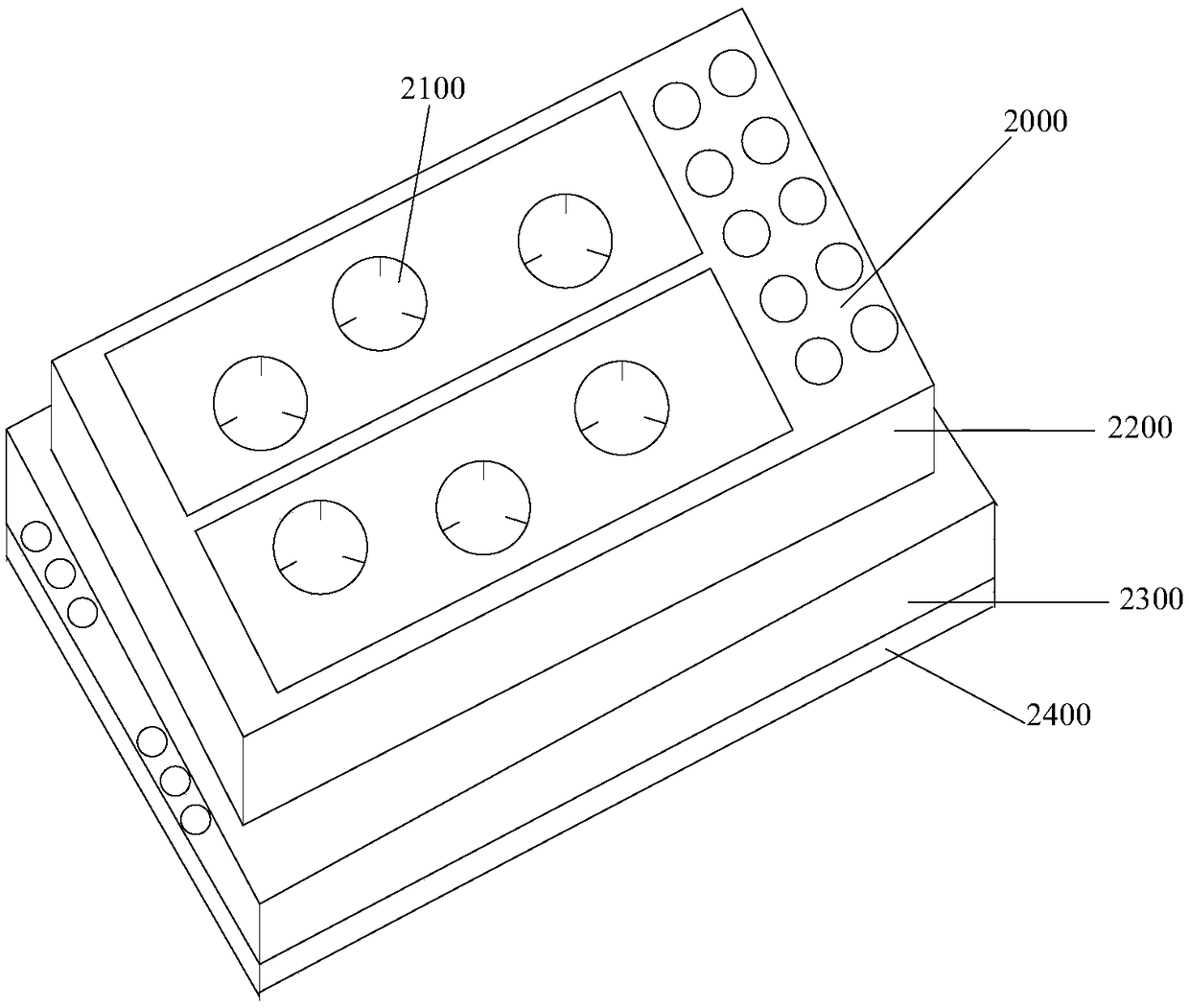
[0117] Step 3. Put the 3D bioprinted tissue containing liver cells and the 3D bioprinted tissue containing tumor cells into the transfer unit 2100, and install the transfer unit 2100 into two different culture chambers on the organ chip 2000;

[0118] Step 4. Put the organ chip 2000 into the connection base 5000, close the base cover 5010 to insert the air guide joint 5042 into the trachea interface;...

example 2

[0134] Step 1. Put the two transfer units 2100 into the two culture chambers on the organ chip 2000 respectively;

[0135] Step 2. Make the printing needle enter the cultivation chamber by adjusting the printing nozzle of the biological 3D printer 1000;

[0136] Step 3. Print the 3D bioprinted tissue with liver cells and the 3D bioprinted tissue with tumor cells on the transfer unit 2100 in the two culture chambers;

[0137] Step 4. Add medium (DMEM medium+10% fetal bovine serum+1% penicillin) to the culture chamber, and perform the operations of steps 4-7 in Example 1;

[0138] Step 5. Cultivate the cells for 20 days, during which the medium is replaced every two days according to step 6 in Example 1;

[0139] Step 6. Execute steps 8-17 in Example 1;

[0140] Step 7. Summarize and analyze the sensing records, cell proliferation results, cell growth state observation records and bio-slice results obtained during the experiment to obtain the hepatocytes and tumor cells on the...

example 3

[0142] Step 1. Execute steps 1-10 in Example 1;

[0143] Step 2. When the concentration of the marker is insufficient or the response speed of the sensor chip is insufficient to cause the medium to flow through, the obtained sensing signal is too low, stop the circulation mode in the drive system 3000, and switch to the alternate mode, set 3 clockwise Pause for 5 seconds after the cycle to perform 2 counterclockwise cycles, the cycle frequency is 0.5Hz, the positive pressure output value is 150kPa, the negative pressure output value is 65kPa, and then the alternate mode is turned on;

[0144] Step 3. Execute steps 9-17 in Example 1, wherein, in step 17, "stop the cycle mode in the drive system 3000" is changed to "stop the alternate mode in the drive system 3000";

[0145] Step 4. Summarize and analyze the sensing records, cell proliferation results, cell growth state observation records and bio-slicing results obtained during the experiment to obtain the hepatocytes and tumor...


PUM


Abstract

The invention provides a system and a method suitable for culturing and real-time monitoring of biological tissue, and relates to the technical field of tissue engineering. The system comprises a biological 3D printer, an organ chip, a connecting base, a drive system and an auxiliary system, wherein the organ chip is connected with the drive system through the connecting base; the biological 3D printer is used for constructing biological 3D printing tissue; the organ chip is used for accommodating a medium and the biological 3D printing tissue and culturing the biological 3D printing tissue; the connecting base is used for accommodating the organ chip and connected with the drive system; the drive system is used for driving the medium to flow in the organ chip; the auxiliary system is usedfor monitoring the state of the biological 3D printing tissue. According to the system and the method, one novel culture platform is provided for works such as tissue culture, pathological research,drug screening and the like.

Description

technical field [0001] The invention relates to the field of tissue engineering, in particular to a system and method suitable for biological tissue culture and real-time monitoring. Background technique [0002] Organ chips provide a new type of culture platform for cell or tissue culture, pathology research, and drug screening. Its main feature is that it can more effectively simulate the interaction of organs in the human body. Organ chips involve three key components, namely living cell tissue / organ components, fluid control components, and detection / sensing components. Fluid control components provide living cell tissue survival substrate and continuous perfusion culture mode, simulating in vivo growth microenvironment; living cell tissue / organ components refer to components that spatially arrange specific cell types in 2D or 3D; detection / Sensing elements provide monitoring and evaluation functions. [0003] Although the 3D tissue structure can better simulate the i...

Claims


Application Information

Patent Timeline
IPC IPC(8): C12M3/00B33Y30/00
CPCC12M33/00B33Y30/00Y02P10/25
Inventor 王玲吕晨泽徐铭恩斯培剑戴嘉韵赖雪聪
Owner REGENOVO BIOTECH
Why Eureka
Try Eureka