Applications of fusion proteins E-cadherin-Fc, VE-cadherin-Fc and VEGF-Fc
A technology of fusion protein and cadherin, applied in biochemical equipment and methods, artificially induced pluripotent cells, embryonic cells, etc., to achieve the effect of improving differentiation efficiency
- Summary
- Abstract
- Description
- Claims
- Application Information
AI Technical Summary
Problems solved by technology
Method used
Image
Examples
Embodiment 1
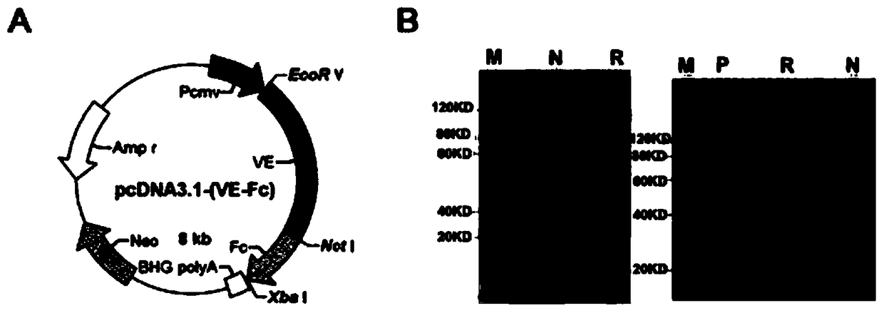
[0093] Embodiment 1: Construct and express human hVE-cad-Fc fusion protein
[0094] See Du Fengyi, PhD dissertation, Nankai University, November 2011. Mainly as follows:
[0095] 1.1 Cloning and sequence analysis of the gene VE-cad in the extracellular domain of vascular endothelial cell cadherin
[0096] According to the human VE-cadherin protein sequence and functional partitions recorded in the UniProt database, specific PCR primers were designed in combination with the gene sequence recorded in GenBank (NCBI Reference Sequence: NM_001795.3) to amplify the hVE-cadherin protein extracellular region (EC1-EC5 ). Upstream primer (P1); 5′-CCG GATATC ATGCAGAGGCTCATGATGCTCC-3' (SEQ ID NO: 1), introduce EcoR V restriction site (underline), downstream primer: (P2) 5'-AA GCGGCCGC TCTGGGCGGCCATATC-3' (SEQ ID NO: 2), a Not I restriction site (underlined) was introduced. Primer synthesis and sequencing were completed by Invitrogen Co., Ltd.
[0097] Extraction of total mRNA from...
Embodiment 2
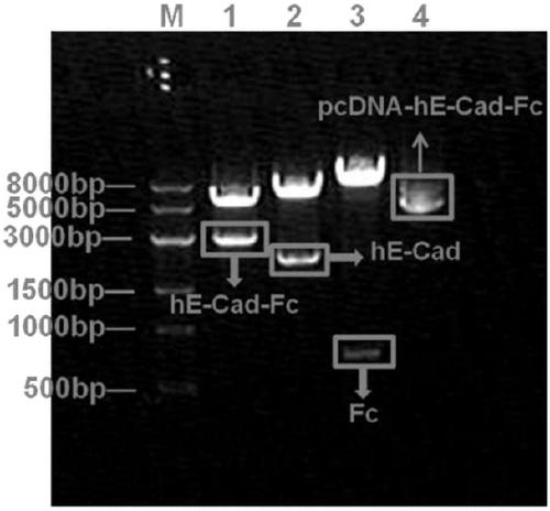
[0126] Embodiment 2: Construct and express human hE-cad-Fc fusion protein
[0127] See Xu Jianbin, PhD dissertation, Nankai University, December 2013. Mainly as follows.
[0128] 2.1 Cloning and sequence analysis of E-cad extracellular domain gene of E-cadherin
[0129] According to the human E-cadherin protein sequence and functional partitions recorded in the UniProt database, specific PCR primers were designed in combination with the gene sequence recorded in GenBank (NCBI Reference Sequence: NM 004360.3) to amplify the extracellular region of the E-cadherin protein. Upstream primer (P1): 5'-CGCAAGCTTATGGGCCCTTG-GAGCCGCAGC-3', SEQ ID NO: 6; Downstream primer: (P2) 5'-TTGCGGCCGCAGGCAGGAATTTGCAATCCTGC-3', SEQ ID NO: 7. Primer synthesis and sequencing were completed by Invitrogen Co., Ltd.
[0130] Total mRNA extraction of L-02 (Cell Bank of Type Culture Collection Committee, Chinese Academy of Sciences): mRNA was extracted according to the conventional method of "Molecular...
Embodiment 3
[0160] Example 3. Detection of hMSC secretion of cytokines and extracellular matrix on the surface of different modified matrices
[0161] Type I collagen (collagen, BD, USA, product number 354249), hE-cad-Fc, hVE-cad-Fc and hE-cad-Fc / hVE-cad-Fc mixed solution ( The ratio of the two fusion proteins is 1:1) were diluted to a final concentration of 10 μg / mL, and 1.5 mL of the diluted collagen solution and hE-cad-Fc / hVE-cad-Fc mixed solution were added to the 6-well cell culture plate , placed in a cell culture incubator at 37° C. for 2 hours, removed and discarded the supernatant, and washed 3 times with 0.01M PBS (pH=7.2).
[0162] followed by a cell density of 10 5 Cells / well hMSCs (Saiye Biotech, China) were inoculated on TCPS culture plates and the culture plates incubated with the above solution, and DMEM / Ham's F12 1: 1 (DF12, BI, USA) culture medium in a cell culture incubator (37°C, 5% CO 2 ) for cultivation. After culturing for 24 h and 48 h respectively, discard the...
PUM
| Property | Measurement | Unit |
|---|---|---|
| particle diameter | aaaaa | aaaaa |
| particle size | aaaaa | aaaaa |
Abstract
Description
Claims
Application Information
 - R&D
- Intellectual Property
- Life Sciences
- Materials
- Tech Scout
- Unparalleled Data Quality
- Higher Quality Content
- 60% Fewer Hallucinations
Browse by: Latest US Patents, China's latest patents, Technical Efficacy Thesaurus, Application Domain, Technology Topic, Popular Technical Reports.
© 2025 PatSnap. All rights reserved.Legal|Privacy policy|Modern Slavery Act Transparency Statement|Sitemap|About US| Contact US: help@patsnap.com


