A nucleic acid aptamer targeting CD133 protein and its screening method and use
A nucleic acid aptamer and targeted combination technology, which is applied in the fields of pharmaceutical formulations, biochemical equipment and methods, medical preparations with non-active ingredients, etc., can solve the problems of undifferentiated thyroid cancer lacking aptamers, etc., and achieve easy Long-term storage, reversible denaturation and renaturation, and easy transportation at room temperature
- Summary
- Abstract
- Description
- Claims
- Application Information
AI Technical Summary
Problems solved by technology
Method used
Image
Examples
Embodiment 1
[0033] Example 1: Screening of nucleic acid aptamers targeting CD133 protein
[0034] a. The aptamer targeting CD133 protein was screened from the ssDNA library by cell screening method;
[0035] b. Detect the ssDNA screened in step a by high-throughput sequencing, and analyze the stability and affinity of the sequence, and screen out a nucleic acid aptamer that targets and binds to the CD133 protein;
[0036] c. Verify that the aptamer targeting CD133 protein selected in step b can specifically target and bind CD133 protein by immunofluorescence method, and express CD133 protein ATC cells combined.
[0037] The sequence of one of the screened nucleic acid aptamers targeting and binding to the CD133 protein is shown as SEQ ID NO.1 in the sequence listing, ie AP-1-M.
Embodiment 2
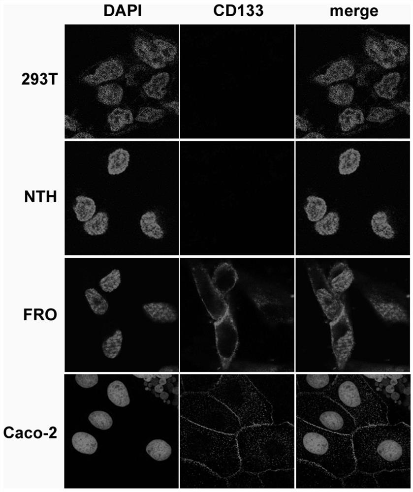
[0038] Example 2: Expression and localization of CD133 protein in different cell lines
[0039] Such as figure 1 Shown, Caco-2: colon adenocarcinoma cell; FRO: undifferentiated thyroid cancer cell; NTH: normal thyroid cell; 293T: human embryonic kidney cell line.
[0040] Confocal Microscopy was used to observe the expression of CD133 protein in different cell lines ( figure 1 ), red fluorescence (ie figure 1The membrane-like distribution of bright spots in the four photomicrographs in the lower left corner of the center) represents the expression distribution of the membrane protein CD133 protein, and the blue fluorescence (ie figure 1 Nucleated bright spots) in each micrograph represent DAPI, indicating cell nuclei. The results showed that CD133 protein was expressed on the cell membrane in undifferentiated thyroid cancer FRO cells and colon adenocarcinoma Caco-2 cells as a positive control group, while HEK-293T cells as a negative control group and normal thyroid cells N...
Embodiment 3
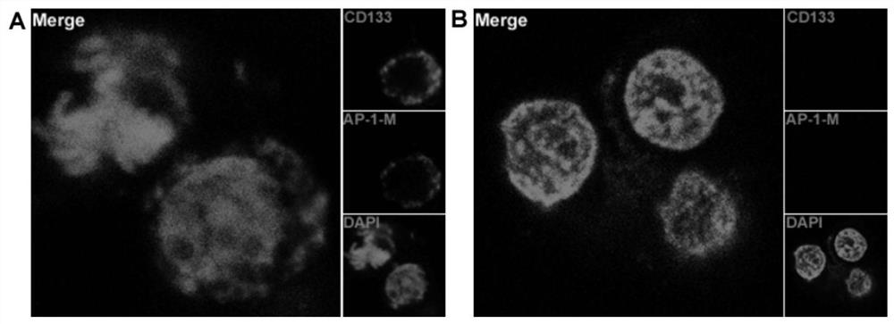
[0041] Example 3: Aptamer AP-1-M binds to CD133 positive cells and negative cells.
[0042] Such as figure 2 As shown, by using FITC to label the AP-1-M nucleic acid aptamer, HEK-293T cells overexpressing CD133 protein were regarded as positive cells, and wild-type HEK-293T cells were regarded as negative cells. The 1-M nucleic acid aptamer was incubated with positive cells and negative cells, and then observed under a laser confocal microscope to find that AP-1-M was combined with positive cells and located on the cell membrane ( figure 2 -A), while not binding to negative cells ( figure 2 -B), which also shows that AP-1-M is a targeting nucleic acid aptamer for CD133 protein.
[0043] The results show that AP-1-M is a nucleic acid aptamer that specifically binds to CD133 protein, and the affinity curve of the nucleic acid aptamer is measured by flow cytometry, and the Kd value of AP-1-M is 101.4nM .
PUM
| Property | Measurement | Unit |
|---|---|---|
| molecular weight | aaaaa | aaaaa |
Abstract
Description
Claims
Application Information
 - R&D
- Intellectual Property
- Life Sciences
- Materials
- Tech Scout
- Unparalleled Data Quality
- Higher Quality Content
- 60% Fewer Hallucinations
Browse by: Latest US Patents, China's latest patents, Technical Efficacy Thesaurus, Application Domain, Technology Topic, Popular Technical Reports.
© 2025 PatSnap. All rights reserved.Legal|Privacy policy|Modern Slavery Act Transparency Statement|Sitemap|About US| Contact US: help@patsnap.com


