Check patentability & draft patents in minutes with Patsnap Eureka AI!

Composition and method for inducing differentiation of mesenchymal stem cells into hypertrophic chondrocytes

A chondrocyte and stem cell technology, applied in the field of stem cells, can solve the problems of low osteogenesis efficiency and less type II collagen in chondrocytes, and achieve the effect of wide application prospect.

Inactive Publication Date: 2019-05-24
SHANGHAI NINTH PEOPLES HOSPITAL AFFILIATED TO SHANGHAI JIAO TONG UNIV SCHOOL OF MEDICINE
View PDF4 Cites 1 Cited by
  • Summary
  • Abstract
  • Description
  • Claims
  • Application Information

AI Technical Summary

Problems solved by technology

[0004] The present invention proposes a composition for inducing the differentiation of mesenchymal stem cells into hypertrophic chondrocytes and its induction method to solve the problem that the cultured chondrocytes lack type II collagen in the prior art, resulting in low osteogenesis efficiency in vivo

Method used


Image

  • Composition and method for inducing differentiation of mesenchymal stem cells into hypertrophic chondrocytes
  • Composition and method for inducing differentiation of mesenchymal stem cells into hypertrophic chondrocytes
  • Composition and method for inducing differentiation of mesenchymal stem cells into hypertrophic chondrocytes

Examples


Embodiment 1

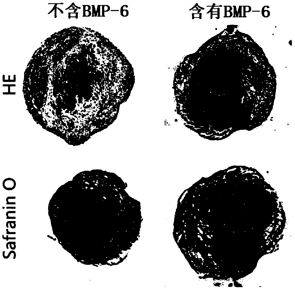
[0035] A composition for inducing differentiation of mesenchymal stem cells into hypertrophic chondrocytes, the culture medium of the chondrogenic stage comprising:

[0036] SFM+dexamethasone (10 -7 mol / L)+ascorbic acid (10 -5 mol / L)+BMP-6(5-20ng / mL)+TGF-β 3 (5-20ng / mL);

[0037] Wherein SFM: DMEM culture fluid+1%HSA+1%PSG+1%HEPES+(0.5-2)%ITS+(0.3-1.2)%linoleic acid.

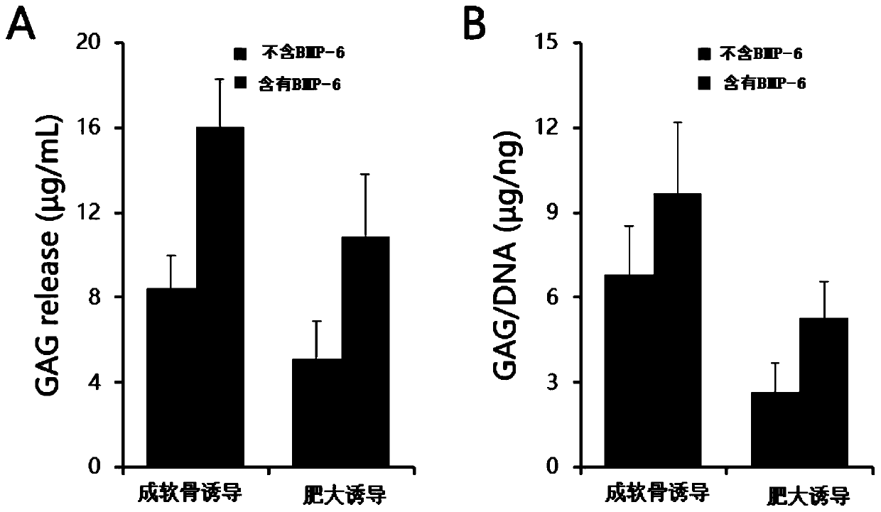
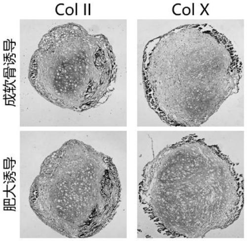
[0038] Media for the hypertrophy induction phase consisted of:

[0039] SFM+SFM+β-glycerophosphate disodium salt (10 -2 mol / L)+dexamethasone (10 -8 mol / L)+ascorbic acid (10 - 5 mol / L);

[0040] Wherein SFM: DMEM culture fluid+1%HSA+1%PSG+1%HEPES+(0.5-2)%ITS+(0.3-1.2)%linoleic acid.

[0041] The culture medium used in this embodiment consists of the following:

[0042] Media for the chondrogenic stage include:

[0043] SFM+dexamethasone (10 -7 mol / L)+ascorbic acid (10 -5 mol / L)+BMP-6(10ng / mL)+TGF-β 3 (10ng / mL);

[0044] Among them, SFM: DMEM culture fluid + 1% HSA + 1% PSG + 1% HEPES + 1% ITS + 0.5...

Embodiment 2

[0064] (1) Preparation of particulate adipose tissue;

[0065] The preparation of particulate adipose tissue is a mature technique in the art, and the steps in this example are as follows:

[0066] (a) Human adipose tissue obtained from liposuction operation, after repeated rinsing with saline, the upper layer of adipose tissue was collected;

[0067] (b) Shred the upper fat tissue with scissors, centrifuge at 1000-2000g for 3-5min, remove the fat in the upper layer and the swelling fluid in the lower layer, and collect the middle fat layer in a 20mL syringe;

[0068] (c) Use a three-way tube to connect two 20mL syringes, inject the two syringes back and forth 30 times, and obtain particulate adipose tissue;

[0069] (d) The particulate adipose tissue is centrifuged at 1000-2000 g for 3-5 minutes to remove the upper layer of fat layer, and the lower layer is the purified particulate adipose tissue.

[0070] (2) In vitro proliferation and culture of particulate adipose tissue...

Embodiment 3

[0089] The culture medium used in this embodiment consists of the following:

[0090] Serum-free basal medium (SFM): DMEM culture solution + 1% HSA + 1% PSG + 1% HEPES + 0.5% ITS + 1.2% linoleic acid; (in order to meet the formal requirements, the end value must be reflected in the implementation, the following same)

[0091] Chondrogenic induction medium: SFM+dexamethasone (10 -7 mol / L)+ascorbic acid (10 -5 mol / L)+BMP-6(20ng / mL)+TGF-β 3 (5ng / mL);

[0092] Hypertrophy induction medium: SFM + β-glycerophosphate disodium salt (10 -2 mol / L)+dexamethasone (10 -8 mol / L)+ascorbic acid (10 -5 mol / L).

[0093] Applying the above medium to the method in Example 1 also meets the requirements.


PUM


Abstract

The invention provides a composition and a method for inducing differentiation of mesenchymal stem cells into hypertrophic chondrocytes. Main compositions in a chondrogenic stage include BMP-6 and TGF-beta 3. The induction scheme of endochondral ossification mainly includes a cartilage induction stage and a hypertrophy induction stage. The differentiation efficiency of the mesenchymal stem cells (MSC) into the chondrocytes is remarkably improved by scientifically proportioning the composition ratios of the BMP-6 and the TGF- beta 3. After the BMP-6 is added, the number of the chondrocytes is obviously increased in HE dye, and the expression of GAG is obviously increased in Safranin O dye. After quantitative GAG detection, the concentration of the GAG in the chondrocytes cultured in culturemedia supplemented with the BMP-6 is obviously higher than that of a control group, and the GAG / DNA content ratio is also obviously increased.

Description

[0001] The invention relates to the technical field of stem cells, in particular to a composition for inducing differentiation of mesenchymal stem cells into hypertrophic chondrocytes and an inducing method thereof. Background technique [0002] The repair of large bone defects is a great challenge for reconstructive surgeons. With more than two million bone grafts performed worldwide each year, it is the second most common tissue transplantation technique after blood transfusion. Among the numerous bone defect treatment methods, autologous bone grafting has been considered the gold standard for bone defect treatment. However, due to the limited donors of autologous bone transplantation and the large damage to the donor site, researchers began to turn to other treatment methods. In recent years, with the development of tissue engineering technology, a small amount of human cells can be used to construct human tissue with biological functions in vitro, which makes people see t...

Claims


Application Information

Patent Timeline
IPC IPC(8): C12N5/077
Inventor 黄如林李青峰王文进李佳奇傅娆
Owner SHANGHAI NINTH PEOPLES HOSPITAL AFFILIATED TO SHANGHAI JIAO TONG UNIV SCHOOL OF MEDICINE
Features
  • R&D
  • Intellectual Property
  • Life Sciences
  • Materials
  • Tech Scout
  • Unparalleled Data Quality
  • Higher Quality Content
  • 60% Fewer Hallucinations
Learn More