
Application of three-dimensional printing technology in establishment of three-dimensional structure digital model in vitro of corneal limbal tissues

A technology of three-dimensional printing and three-dimensional structure, which is applied in the field of corneal anatomy and can solve the problems of simulating the three-dimensional digital structure of limbal tissue and so on.

Active Publication Date: 2019-06-07
TONGJI HOSPITAL ATTACHED TO TONGJI MEDICAL COLLEGE HUAZHONG SCI TECH
View PDF6 Cites 1 Cited by
  • Summary
  • Abstract
  • Description
  • Claims
  • Application Information

AI Technical Summary

Problems solved by technology

[0004] At present, there is no technology that can simulate the three-dimensional digital structure of limbal tissue in vitro

Method used


Image

  • Application of three-dimensional printing technology in establishment of three-dimensional structure digital model in vitro of corneal limbal tissues
  • Application of three-dimensional printing technology in establishment of three-dimensional structure digital model in vitro of corneal limbal tissues

Examples


Embodiment Construction

[0036] The present invention will be described in further detail below in conjunction with specific examples, so that those skilled in the art can understand, but do not limit the implementation of the present invention, other reagents and equipment well known in the art are applicable to the implementation of the following schemes of the present invention.

[0037] The equipment and materials that the present invention needs have: operating microscope, microsurgical instrument, paraffin slicer, all are provided by purchasing in the market; Optical microscope is Xiamen Motic BA600 type, is equipped with Moticam microscope camera and Motic digital slice scanning and application system software.

[0038] Preparation of reagents and working solutions involved in the present invention: the fixative is composed of 60mL of absolute ethanol, 10mL of neutral formaldehyde with a volume fraction of 10%, 10mL of glacial acetic acid, and 20mL of chloroform. Xylene, hematoxylin, eosin, and ...


PUM


Abstract

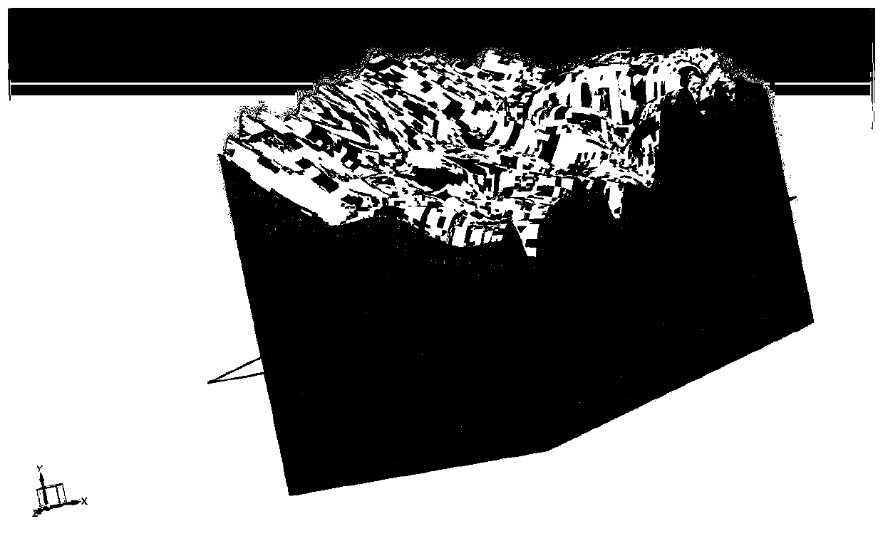
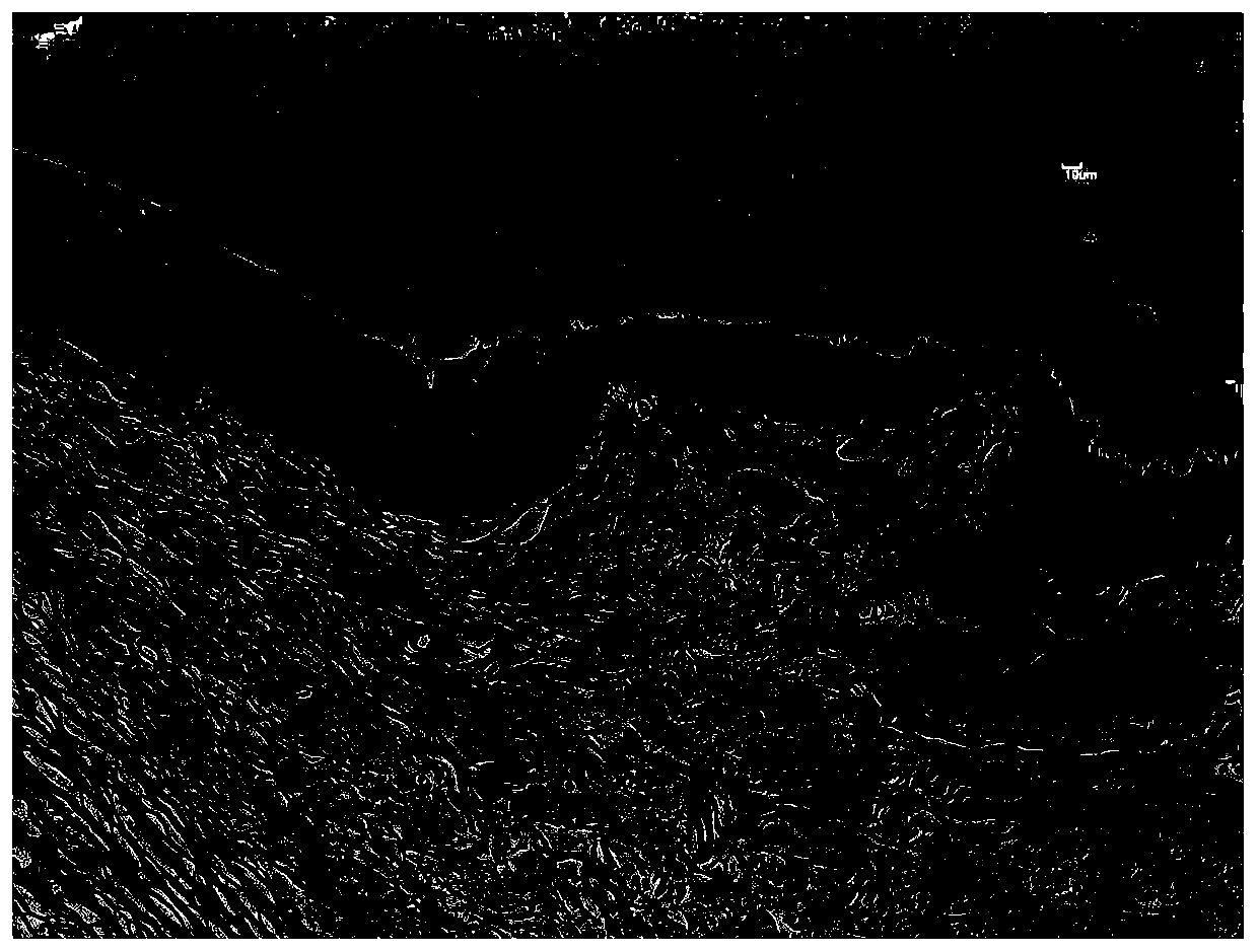
The invention belongs to the technical field of corneal anatomy, and particularly discloses an application of a three-dimensional printing technology in corneal limbal tissue in-vitro three-dimensional modeling, and the method of the application comprises the steps of specimen fixation, tissue embedding and slicing, HE staining, image acquisition, digitization and three-dimensional reconstructionto obtain a three-dimensional structure digitized model of corneal limbal tissue. The reconstructed corneal limbal three-dimensional stereogram can display the microstructure of the human corneal limbal tissue, and therefore it is indicated that the method for conducting three-dimensional digital reconstruction by collecting the high-definition microscopic tissue image obtained after slice dyeingis feasible. The method combines the advantages that adjacent tissue images are easy to align under the low-power lens and tissue images under the high-power lens are clearer, is simple and practical,and can also be used for microstructure three-dimensional reconstruction of other tissues.

Description

technical field [0001] The invention relates to the technical field of corneal anatomy, in particular to the application of a three-dimensional printing technology in three-dimensional modeling of limbal tissue in vitro. Background technique [0002] The corneal limbus is the transition zone between the cornea and the sclera, about 1 mm wide, and its anatomical structure consists of epithelial cell layer and stroma layer from top to bottom. The epithelial cell layer is more than 10 layers of irregularly arranged small cylindrical cells, and part of the epithelial cells at the base protrude downwards into the matrix, while the matrix extends into the epithelial layer in a finger-like manner, forming a staggered arrangement of epithelial-matrix Vogt Palisades. Limbal stem cells are distributed in the basal cells of the Vogt palisade structure. They are adult stem cells with the ability to self-renew and differentiate into corneal epithelial cells. It is an important guarantee...

Claims


Application Information

Patent Timeline
Patent Type & Authority Applications(China)
IPC IPC(8): G06T17/00
CPCY02P10/25
Inventor 王玮李贵刚徐玲娟李阳王云明
Owner TONGJI HOSPITAL ATTACHED TO TONGJI MEDICAL COLLEGE HUAZHONG SCI TECH
Learn More