Preparation method of hydrogel porous microspheres and porous scaffold material

A technology of hydrogel microspheres and porous microspheres is applied in the field of tissue engineering materials, which can solve the problems of uncontrollable and stable formation of spheres, and achieve the effect of maintaining cell viability.

Inactive Publication Date: 2019-06-14
上海市伤骨科研究所
View PDF5 Cites 22 Cited by
  • Summary
  • Abstract
  • Description
  • Claims
  • Application Information

AI Technical Summary

Problems solved by technology

This method is simple to operate and does not require special equipment, but the state of ball formation is uncontrollable, and stable and uniform microspheres cannot be formed.

Method used


Image

  • Preparation method of hydrogel porous microspheres and porous scaffold material
  • Preparation method of hydrogel porous microspheres and porous scaffold material
  • Preparation method of hydrogel porous microspheres and porous scaffold material

Examples


preparation example Construction

[0042] According to different hydrogel materials, this application also provides a method for preparing porous hydrogel microspheres, which includes the following steps:

[0043] A) Mixing the hydrogel material and the buffer to obtain a hydrogel solution; mixing the oily material and the surfactant to obtain the oily solution;

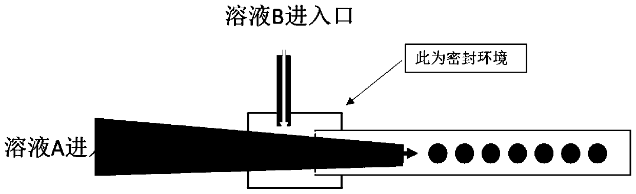
[0044] B) The first container is partially nested into the second container, the inner diameter of the inlet end of the first container is greater than the inner diameter of the outlet end, and the inner diameter of the outlet end of the first container is smaller than the inner diameter of the second container; The hydrogel solution is injected into the first container, the oily solution is injected into the intersection of the first container and the second container, and the oily solution and the hydrogel solution are under the action of flow shearing force. Form water-in-oil droplets;

[0045] C) Cross-linking the water-in-oil droplets to obtain hydroge...

Embodiment 1

[0055] 1) Solution A: Add 2ml of phosphate buffer solution to 100mg of methacrylic group-modified gelatin, and add 20mg of photoinitiator at the same time, place it in a 60°C water bath for 45 minutes, shake it up intermittently, and fully dissolve it. The solution becomes colorless, clear and transparent for use;

[0056] 2) Solution B: Take 50ml of perfluorinated oil and add 1% by mass to the surfactant span80, shake to dissolve, and remove bubbles by ultrasound;

[0057] 3) Capillary glass tube C: Take a capillary glass tube with an inner diameter of 100 μm, draw a wire at the distal end, and cut a capillary with an outlet inner diameter of 50 μm under a microscope;

[0058] 4) Capillary glass tube D: Capillary glass tube with an inner diameter of 100μm;

[0059] 5) Sleeve the outlet end of tube C into tube D, place it on a glass slide, and fix it with gum; use a 21G syringe needle nest to close the junction of tube C and tube D, and at the same time nest a 21G syringe needle at th...

Embodiment 2

[0073] 1) Solution A: Add 2ml of deionized water to 80mg of methacrylic group-modified gelatin, then add 20mg of sodium alginate, and add 20mg of photoinitiator at the same time, place it in a 60℃ water bath for 45 minutes with intermittent shaking Evenly, fully dissolve, wait for the solution to become colorless, clear and transparent;

[0074] 2) Solution B: Take 50ml of perfluorinated oil and add 1% by mass to the surfactant span80, shake to dissolve, and remove bubbles by ultrasound;

[0075] 3) Capillary glass tube C: Take a capillary glass tube with an inner diameter of 100 μm, draw a wire at the distal end, and cut a capillary with an outlet inner diameter of 50 μm under a microscope;

[0076] 4) Capillary glass tube D: Capillary glass tube with an inner diameter of 100μm;

[0077] 5) Put the outlet end of tube C into tube D, place it on a glass slide, fix it with gum, and close the junction of tube C and tube D with a 21G syringe needle nesting, and at the same time nest a 21G...


PUM

PropertyMeasurementUnit
pore sizeaaaaaaaaaa
particle diameteraaaaaaaaaa
pore sizeaaaaaaaaaa

Abstract

The invention provides a preparation method of hydrogel porous microspheres. The preparation method comprises the following steps: a, a hydrogel material, a buffer solution and a photoinitiator are mixed to obtain a hydrogel solution, and an oily material is mixed with a surfactant to obtain an oily solution; b, a first container is partially nested in a second container, the inner diameter of theinlet end of the first container is larger than the inner diameter of the outlet end of the first container, and the inner diameter of the outlet end of the first container is smaller than the innerdiameter of the second container; the hydrogel solution is injected into the first container, the oily material is injected into the intersection of the first container and the second container, and the oily solution and the hydrogel solution form water-in-oil droplets under the action of a flowing shear force; c, ultraviolet irradiation is performed on the water-in-oil droplets, and crosslinkingis performed to obtain hydrogel microspheres; and d, freezing and freeze-drying are performed on the hydrogel microspheres to obtain the hydrogel porous microspheres. According to the invention, uniform and stable hydrogel porous microspheres are obtained by utilizing a micro-fluidic technology.

Description

Technical field [0001] The invention relates to tissue engineering materials, in particular to a preparation method of hydrogel porous microspheres and porous scaffold materials. Background technique [0002] The non-renewable necrosis or loss of local tissues caused by disease, trauma or injury in the human body is a major problem in the current medical field. These tissues often need to be replaced or transplanted for regeneration. However, autologous transplantation or allogeneic transplantation treatment has major problems. First, the graft is expensive, the medical burden is heavy, and the patient is painful. At the same time, there is often rejection of immune tissue during the transplantation process, resulting in failure of the operation. [0003] Tissue engineering technology aims to maintain or improve patient functions through the development of biological alternative materials, and has important application prospects. With the development of tissue engineering technolo...

Claims


Application Information

Patent Timeline
Patent Type & Authority Applications(China)
IPC IPC(8): C08J9/28C08J3/075C08J3/24C08L89/00C08L5/04C08K3/32B01J13/14A61L27/56A61L27/52A61L27/54A61L27/50A61L27/04A61L27/10A61L27/38A61L27/22
Inventor 崔文国
Owner 上海市伤骨科研究所
Why Eureka
Try Eureka