Medical imaging method and photon counting energy spectrum CT imaging device
A photon counting and medical imaging technology, applied in the field of medical imaging, can solve the problems of difficult data link design, high hardware design requirements, affecting image quality, etc., to reduce the amount of scanning data, meet the actual clinical needs, and ensure the spatial resolution of images. rate effect
- Summary
- Abstract
- Description
- Claims
- Application Information
AI Technical Summary
Problems solved by technology
Method used
Image
Examples
Embodiment 1
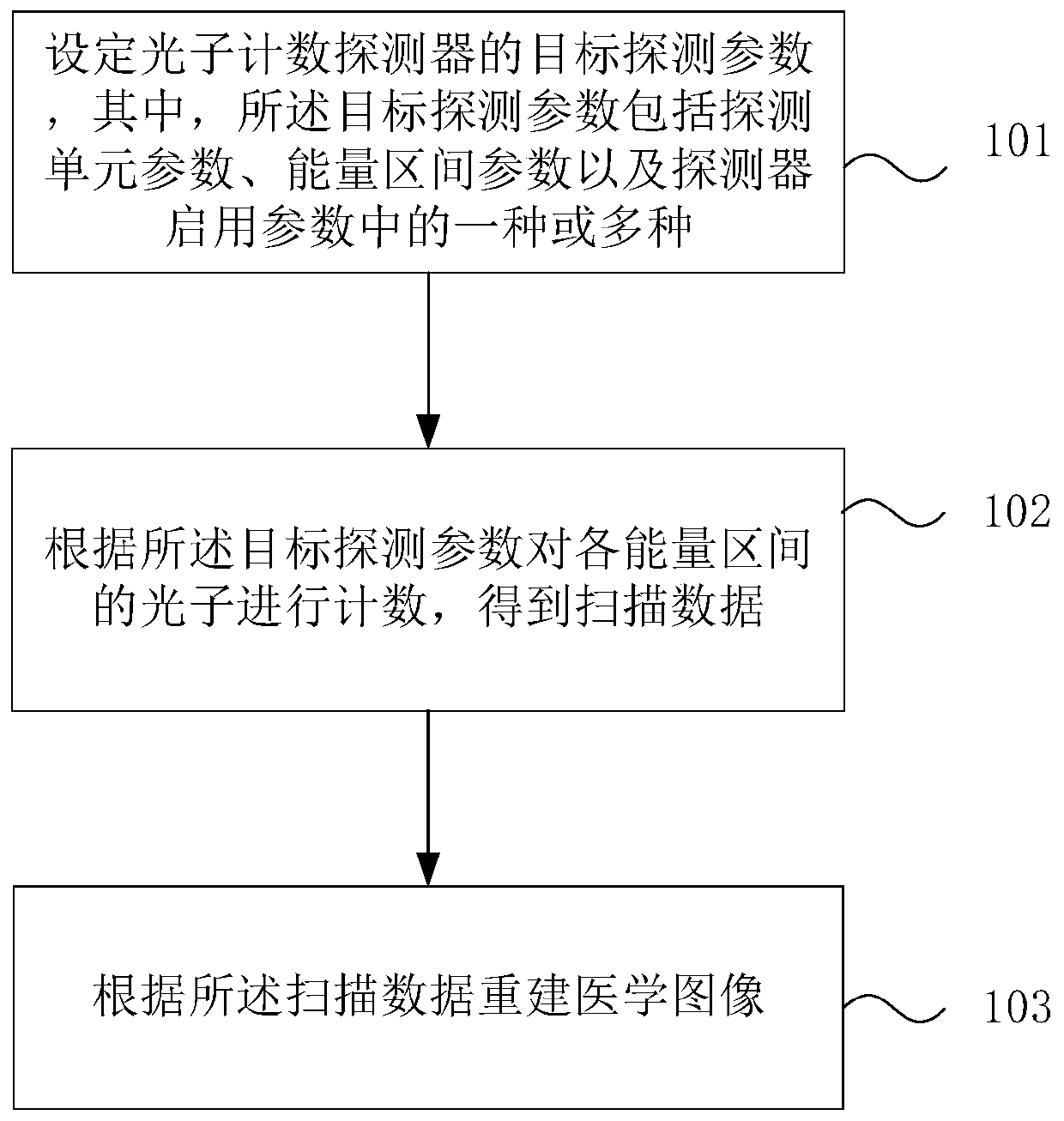
[0022] figure 1 It is a flow chart of the medical imaging method provided by Embodiment 1 of the present invention. This embodiment is applicable to imaging of medical equipment, especially applicable to imaging of photon counting spectral CT imaging equipment. The method can be performed by a photon counting energy spectrum CT imaging device, the device can be implemented by hardware and / or software, and the device can be integrated into a device (such as a photon counting energy spectrum CT imaging device) for execution, and specifically includes the following steps :
[0023] Step 101, setting the target detection parameters of the photon counting detector.
[0024] Wherein, the target detection parameters include one or more of detection unit parameters, energy interval parameters, and detector enabling parameters.
[0025] Among them, the photon counting detector has energy discrimination capability and is equipped with an application-specific integrated circuit. The X-...
Embodiment 2
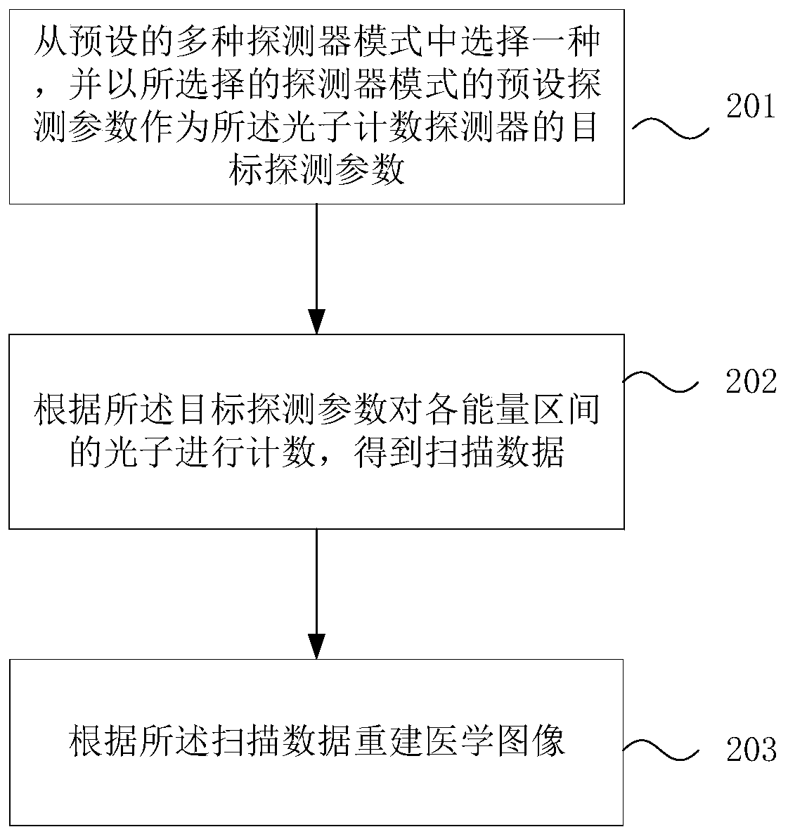
[0045] Figure 2a It is a flowchart of a medical imaging method provided by Embodiment 2 of the present invention. On the basis of the above embodiments, in this embodiment, optionally, the setting of the target detection parameters of the photon counting detector includes: Select one of the multiple detector modes, and use the preset detection parameters of the selected detector mode as the target detection parameters of the photon counting detector.
[0046] On this basis, further, the detector mode includes a first mode and a second mode, wherein the detector counting unit size of the first mode is smaller than the detector counting unit size of the second mode.
[0047] On this basis, further, the number of energy intervals in the first mode is lower than the number of energy intervals in the second mode.
[0048] On this basis, further, the number of pixel rows enabled in the photon counting detector in the first mode is smaller than the number of pixel rows enabled in t...
Embodiment 3
[0080] image 3 A schematic structural diagram of a photon counting energy spectrum CT imaging device provided in Embodiment 3 of the present invention, as shown in image 3 As shown, the device includes a radiation source 31 , a photon counting detector 32 and a detector control unit 33 .
[0081] Wherein, the ray source 31 is used to generate X-ray photons;
[0082] A photon counting detector 32, comprising a plurality of detector pixels, is used to generate scan data according to detection of X-ray photons passing through the target object; the photon counting detector can divide one or more energy intervals according to the energy of the photons, and Counting the detected photons in each energy interval; the photon counting detector is configured to adjust detection parameters;
[0083] The detector control unit 33 is configured to configure detection parameters of the photon counting detector according to instructions.
[0084] Optionally, configuring the detection param...
PUM
 Abstract
Description
Claims
Application Information
 - R&D
- Intellectual Property
- Life Sciences
- Materials
- Tech Scout
- Unparalleled Data Quality
- Higher Quality Content
- 60% Fewer Hallucinations
Browse by: Latest US Patents, China's latest patents, Technical Efficacy Thesaurus, Application Domain, Technology Topic, Popular Technical Reports.
© 2025 PatSnap. All rights reserved.Legal|Privacy policy|Modern Slavery Act Transparency Statement|Sitemap|About US| Contact US: help@patsnap.com


