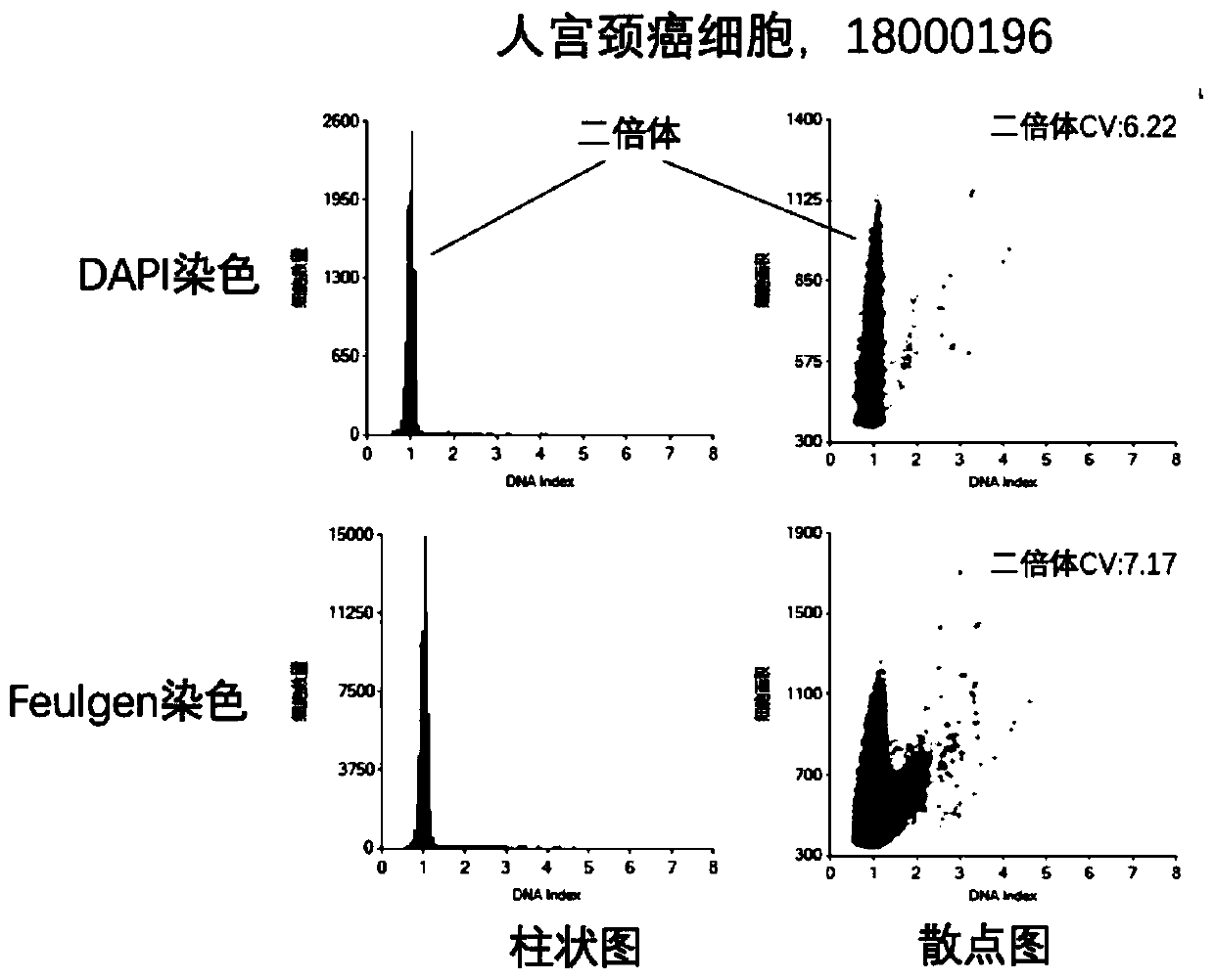
Cancer screening and diagnosis method via staining quantification of cellular DNA by fluorescent dye DAPI
A fluorescent dye and cell technology, applied in the field of DNA detection, can solve the problems of long time, low quantitative accuracy, complicated dyeing operation, etc., and achieve the effect of simple operation, accurate quantification, and fast dyeing time
- Summary
- Abstract
- Description
- Claims
- Application Information
AI Technical Summary
Problems solved by technology
Method used
Image
Examples
Embodiment 1
[0024] Embodiment one, real-time staining, comprises the following steps:
[0025] Step 1: Use a cervical brush to brush some cells from the female cervix, place the cervical brush in a fixative solution containing alcohol and methanol, and shake the cell sample for 1 min to suspend it.
[0026] Step 2, take the cell sedimentation chamber method to make slices: take the filter, add 2.5ml of cell separation solution, and cover the filter;
[0027] Step 3, take 20-100ul DAPI 10* working solution and 300ul cell samples, mix evenly for 1 minute, and stain for 3 minutes;
[0028] Step 4, transfer the sample to the production chamber;
[0029] Step 5, centrifuge, 3min;
[0030] Step 6, take slides, wash with water for 20s, and wash with PBS for 30s×3 times;
[0031] Step 7, dehydration with 95% and 100% ethanol for 1 min each;
[0032] Step 8, wait until dry, and seal with resin;
[0033] Step 9: Examine under a fluorescent microscope for quantitative analysis.
[0034] Step 1...
Embodiment 2
[0036] Embodiment 2, integration of fixation and dyeing, comprises the following steps:
[0037] Step 1, improve the cell fixative and add DAPI working solution to make the final concentration between (0.2-1.0ug / ml).
[0038] Step 2, use a cervical brush to brush some cells from the female cervix, place the cervical brush in the fixative solution containing DAPI, and shake the cell sample for 2 minutes to make it suspended.
[0039] Step 3, add 2.5ml of cell separation medium to the cell sedimentation chamber, and cover the filter;
[0040] Step 4, take a 300ul cell sample and transfer the sample to the production chamber;
[0041] Step 5, centrifuge, 3min;
[0042] Step 6, take slides, wash with water for 20s, and wash with PBS for 30s×3 times;
[0043] Step 7, 95% and 100% ethanol dehydration for 1 min each;
[0044] Step 8, wait until dry, and seal with resin;
[0045] Step 9, check under a fluorescent microscope for quantitative analysis;
[0046] Step 10, identifyin...
Embodiment 3
[0047] Embodiment three, paraffin section, comprises the following steps:
[0048] Step 1, generally use a constant temperature water bath, the temperature is controlled at about 37-40°C, which is conducive to the unfolding of paraffin sections, and the pasted slices are dried in a 60°C constant temperature box for 2 hours, and the protein can be stained after solidification.
[0049] Step 2, dewaxing and hydration of the slices: the dried slices were dewaxed in xylene, and then dewaxed with pure alcohol, 95%, 85%, and 70% in descending order from high concentration to low concentration. change.
[0050] Step 3, soak the slices in 2ug / ml DAPI staining solution for 10 minutes.
[0051] Step 4, take slides, wash with water for 20s, and wash with PBS for 30s×3 times;
[0052] Step 5, dehydration with 95% and 100% ethanol for 1 min each;
[0053] Step 6, wait until dry, and seal with resin;
[0054] Step 7, check under a fluorescent microscope for quantitative analysis;
[0055...
PUM
 Abstract
Description
Claims
Application Information
 - R&D
- Intellectual Property
- Life Sciences
- Materials
- Tech Scout
- Unparalleled Data Quality
- Higher Quality Content
- 60% Fewer Hallucinations
Browse by: Latest US Patents, China's latest patents, Technical Efficacy Thesaurus, Application Domain, Technology Topic, Popular Technical Reports.
© 2025 PatSnap. All rights reserved.Legal|Privacy policy|Modern Slavery Act Transparency Statement|Sitemap|About US| Contact US: help@patsnap.com


