Coating buffer and separation culture method of primary tumor cells

A technology for primary tumor cells and cell culture, which is applied in the field of coating solution to promote cell adhesion and the separation and culture of primary tumor cells, which can solve the problems of the complex process of primary tumor cell culture, slow adherence speed, and small number of cells, etc. question

Active Publication Date: 2019-07-26
BEIJING HARMONY HEALTH MEDICAL DIAGNOSTICS CO LTD
View PDF3 Cites 11 Cited by
  • Summary
  • Abstract
  • Description
  • Claims
  • Application Information

AI Technical Summary

Problems solved by technology

[0004] Because the culture process of primary tumor cells is too complicated, the speed of adherence is slow, the number of cells is small, the purity is low, and the culture is difficult, etc., the existing culture methods of primary tumor cells often need to use relatively large tissue samples for culture.
In many cases, the tissue samples obtained cannot meet the needs of the existing culture methods, that is, for tumor cell tissues with the characteristics of small tissue samples, mixed fibroblasts, and microbial diversity, such as intestinal cancer cells, gastric cancer, etc. For cell tissues, etc., some existing primary tumor cell culture media are difficult to use to cultivate primary tumor cells

Method used


Image

  • Coating buffer and separation culture method of primary tumor cells
  • Coating buffer and separation culture method of primary tumor cells
  • Coating buffer and separation culture method of primary tumor cells

Examples


Embodiment 1

[0098] Example 1: Isolation and culture of primary intestinal cancer cells 1

[0099] (1) Evenly spread the coating solution used to promote cell adhesion on 25cm 2 In the cell culture bottle, place it horizontally in a cell culture incubator at 37°C for 1-2 hours, wait until the liquid in the culture bottle is completely solidified, and obtain a coated culture bottle for later use.

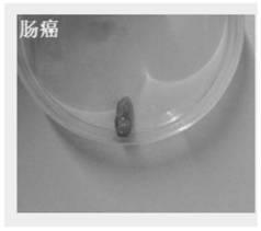
[0100] (2) Will obtain such as figure 2 0.3 g of the fresh intestinal cancer tissue indicated was transferred to a cell culture dish, and treated with 500 μg / mL penicillin, 100 μg / mL kanamycin sulfate, 0.5 μg / mL amphotericin B, 3 μg / mL vancomycin, 5 μg Rinse with normal saline of cefmetazole sodium 5-8 times to remove fat, mucous membrane and other non-cancerous tissue impurities.

[0101] (3) The intestinal cancer tissue treated in (2) was transferred to a new culture dish, and the tissue block was chopped into fine pieces with scissors and blades.

[0102] (4) Transfer the minced tumor tiss...

Embodiment 2

[0109] Example 2: Isolation and culture of primary intestinal cancer cells 2

[0110] (1) Evenly spread the coating solution used to promote cell adhesion on 25cm 2 In the cell culture bottle, place it horizontally in a cell culture incubator at 37°C for 1-2 hours, wait until the liquid in the culture bottle is completely solidified, and obtain a coated culture bottle for later use.

[0111] (2) Transfer the obtained fresh intestinal cancer tissue to a cell culture dish, and mix with 500 μg / mL penicillin, 500 μg / mL kanamycin sulfate, 0.25 μg / mL amphotericin B, 0.5 μg / mL vancomycin, Rinse with 5 μg / mL cefmetazole sodium saline 5-8 times to remove non-cancerous tissue impurities such as fat and mucous membrane.

[0112] (3) The intestinal cancer tissue treated in (2) was transferred to a new culture dish, and the tissue block was chopped into fine pieces with scissors and blades.

[0113](4) Transfer the minced tumor tissue in (3) to a 50mL centrifuge tube, resuspend in 10-20m...

Embodiment 3

[0120] Example 3: Isolation and culture of primary intestinal cancer cells 3

[0121] (1) Evenly spread the coating solution used to promote cell adhesion on 25cm 2 In the cell culture bottle, place it horizontally in a cell culture incubator at 37°C for 1-2 hours, wait until the liquid in the culture bottle is completely solidified, and obtain a coated culture bottle for later use.

[0122] (2) Transfer the obtained fresh intestinal cancer tissue to a cell culture dish, and use 20 μg / mL penicillin, 20 μg / mL kanamycin sulfate, 0.25 μg / mL amphotericin B, 2 μg / mL vancomycin, 5 μg / mL cefmetazole sodium saline was washed 8 times to remove non-cancerous tissue impurities such as fat and mucous membrane.

[0123] (3) The intestinal cancer tissue treated in (2) was transferred to a new culture dish, and the tissue block was chopped into fine pieces with scissors and blades.

[0124] (4) Transfer the minced tumor tissue in (3) to a 50mL centrifuge tube, resuspend in 10-20ml DF medi...


PUM


Abstract

The invention provides a coating buffer for promoting cell adherence and a separation culture method of primary tumor cells. The coating buffer for promoting cell adherence comprises a type I collagen, a coating solution II and a coating solution III which are uniformly mixed, wherein the coating buffer II is a mixture containing 10*F12 medium, and the coating buffer III is a mixture composed of NaOH, NaHCO3 and HEPES. The scheme achieves separation culture of the primary tumor cells from smaller tissue samples.

Description

technical field [0001] The invention relates to the technical field of in vitro cell culture, in particular to a coating solution for promoting cell adhesion and a method for separating and culturing primary tumor cells. Background technique [0002] With the improvement of people's living standards, the number of cancer patients is increasing, and the number of cancer patients tends to be younger. In order to study its pathogenesis and treatment, tumor cell lines of different tumors have been established. However, after long-term culture, the biological specificity of tumor cell lines has changed, which is not conducive to the study of its carcinogenesis, molecular genetics, immunological characteristics, and evolution mechanism of invasion and metastasis. [0003] For the primary cultured tumor cells, their biological characteristics have not changed much, and they still retain the original diploid heredity, and the gene retention is more than 90%, which is suitable for d...

Claims


Application Information

Patent Timeline
Patent Type & Authority Applications(China)
IPC IPC(8): C12N5/09
CPCC12N5/0693C12N2509/00
Inventor 智慧芳佟洪梅贾玉霞倪君君
Owner BEIJING HARMONY HEALTH MEDICAL DIAGNOSTICS CO LTD
Why Eureka
Try Eureka