Biological tissue transparence imaging technology
A biological tissue and transparent technology, applied in the field of biological sample processing and optical imaging, can solve the problems of unfavorable clearing time, fluorescent protein retention, long operation time, high fat content, etc., to improve the clearing efficiency, shorten the required time, and quickly The effect of transparency
- Summary
- Abstract
- Description
- Claims
- Application Information
AI Technical Summary
Problems solved by technology
Method used
Image
Examples
Embodiment 1
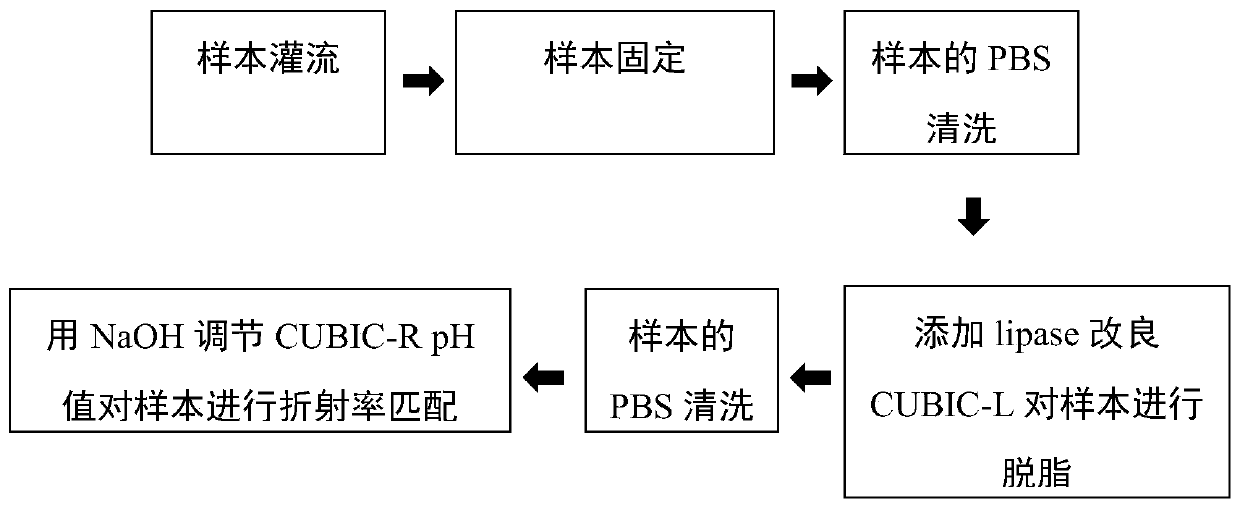
[0059] This embodiment provides a biological tissue transparent imaging method, which is an improvement on the CUBIC optical transparency scheme.
[0060] For the optical imaging application of CUBIC light-clearing technology in high-fat liver samples, we first optimized the protocol of CUBIC light-clearing technology. The sample used was the liver lobe of 8-week-old CX3CR1-mTmG mice. The steps included:
[0061] (1) Preparation of 1×PBS: Prepare 1L of 1×PBS with PBS powder, adjust the pH value to 7.4 with 1mol NaOH solution, then filter the prepared PBS with a filter membrane and store it at 4°C for later use.
[0062] (2) Preparation of 4% PFA: Prepare 4% PFA with prepared 1×PBS, adjust the pH value to 7.4, and store at 4°C.
[0063] (3) Prepare CUBIC-L reagent: 10% mass volume ratio of TritonX-100 and 10% mass volume ratio of N-butylethylenediamine, such as 40g TritonX-100 and 40g N-butylethylenediamine to constant volume Dissolve to 400mL ddH 2 O middle. Use a micromete...
Embodiment 2-4
[0074] Embodiments 2-4 respectively provide a biological tissue transparent imaging method, and the steps of each method are basically the same as those in Embodiment 1, and the differences are as follows:
[0075] In Example 2, the preparation method of CUBIC-L reagent A is: use a micrometer balance to weigh 100-500U / mg lipase powder and dissolve it into the prepared CUBIC-L, so that the lipase content reaches 4-20U / mL. All the other steps are consistent with Example 1.
[0076] In Example 3, after normal perfusion was performed on the dead mice, the PBS reagent containing saturated lipase was slowly perfused for 1 hour. The method of preparing lipase-containing PBS saturated reagent is as follows: add 0.4g 100-500U / mg lipase powder to 200mL 1×PBS, dissolve and mix well, filter with 200 / 400 mesh gauze to remove the insoluble part, and store at 4°C. Both CUBIC-L reagent A and CUBIC-L reagent B do not contain lipase, that is, the CUBIC-L reagent prepared in the same way as in ...
Embodiment 5
[0080] This example provides the optical imaging effect of the sample treated with the improved CUBIC scheme by adding lipase.
[0081] In order to detect the effect of the CUBIC protocol on fluorescence retention under different improved conditions, we performed confocal microscopic optical imaging on the liver lobes of CX3CR1-mTmG mice after clearing in Example 1 and Example 2:
[0082] We used the 10x water lens of LSM 780NLO confocal microscope (Zeiss, Germany) for imaging, and the excitation light was 488nm and 633nm. Under the same imaging conditions, different groups of samples were imaged fluorescently. Figure 5 It can be seen that each group is relatively good at maintaining RFP fluorescence, and the imaging depth can reach more than 2.6mm. However, in terms of the uniformity of RFP fluorescence retention of samples, the 4-20U / mL group has the best effect; for GFP As for the retention of fluorescence, it can be clearly seen that compared with the high-concentration l...
PUM
 Abstract
Description
Claims
Application Information
 - R&D
- Intellectual Property
- Life Sciences
- Materials
- Tech Scout
- Unparalleled Data Quality
- Higher Quality Content
- 60% Fewer Hallucinations
Browse by: Latest US Patents, China's latest patents, Technical Efficacy Thesaurus, Application Domain, Technology Topic, Popular Technical Reports.
© 2025 PatSnap. All rights reserved.Legal|Privacy policy|Modern Slavery Act Transparency Statement|Sitemap|About US| Contact US: help@patsnap.com


