

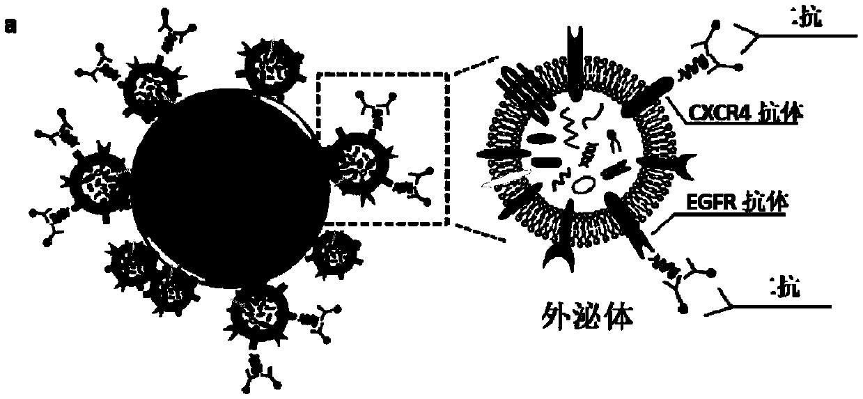
Exosome detecting device for detection and by-stage judgment of non-small-cell lung cancer and application
A non-small cell lung cancer and detection device technology, applied in the field of medical detection, can solve problems such as difficulty
- Summary
- Abstract
- Description
- Claims
- Application Information
AI Technical Summary
Problems solved by technology
Method used
Image

Examples
Embodiment 1
[0107] This example is used to illustrate the extraction of exosomes from the cell culture supernatant.
[0108] 1. Cell material used
[0109] Colorectal cancer cell line SW620, three lung cancer cell lines A549, H1975, H1650.
[0110] 2. The specific steps include:
[0111] 1) When the cells grow to a density of 60%, replace with the culture medium prepared from the serum that has previously deducted exosomes, and place it in the cell culture incubator for 48 hours.
[0112] Wherein, the preparation of the culture medium by deducting the serum of exosomes comprises the following steps:
[0113] ① Add the serum to the basal medium, so that the concentration of FBS in the cell culture medium is 20%;
[0114] ②Ultracentrifuge the cell culture medium overnight at 4°C with a centrifugal force of 100,000 g;
[0115] ③ After the ultracentrifugation, collect the supernatant, dilute the medium containing 20% FBS with the basal medium to a FBS concentration of 10%, and add 1% do...
Embodiment 2
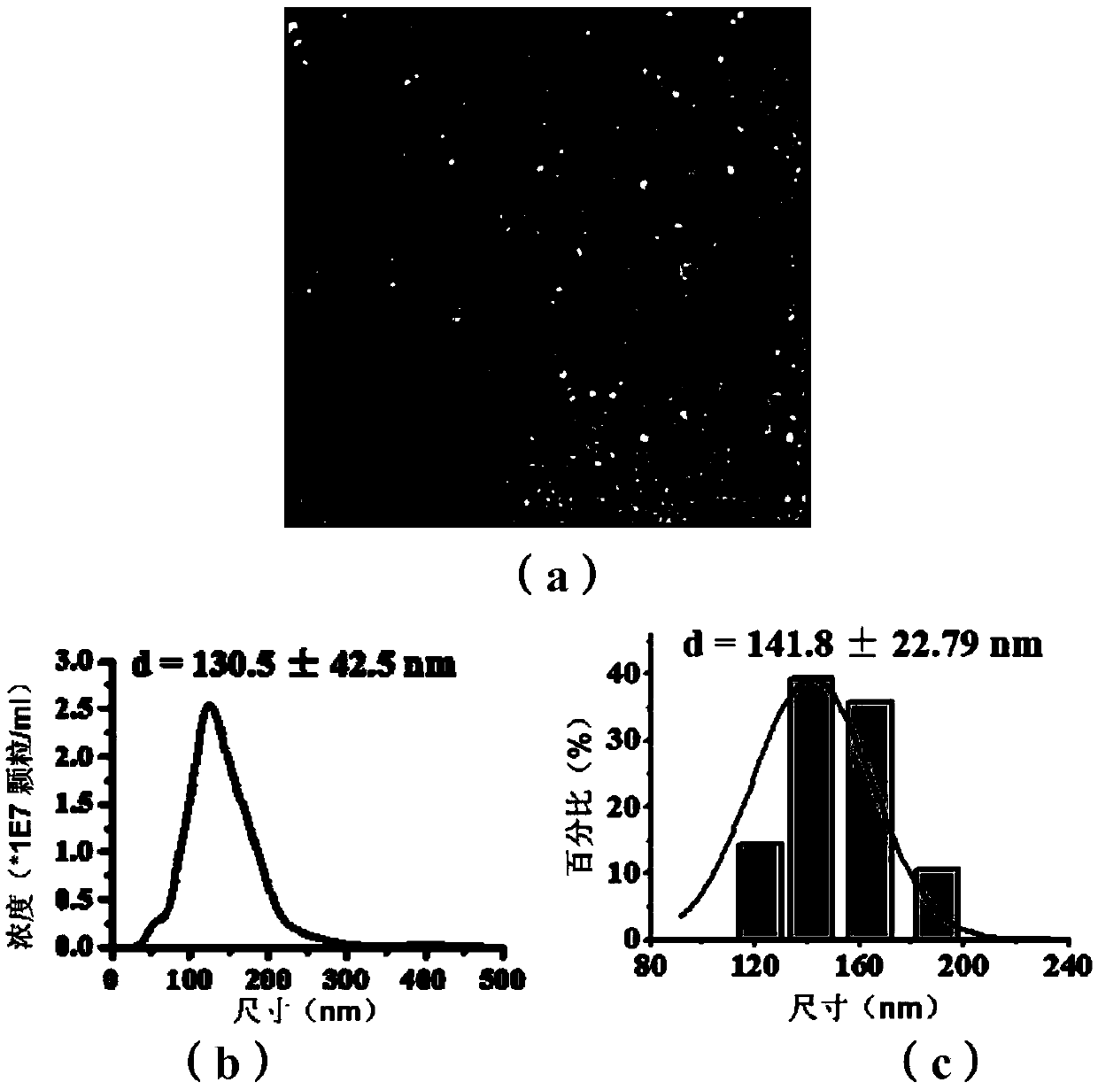
[0123] This example is used to illustrate the morphology and size characterization of the extracted exosomes.
[0124] 1. Transmission Electron Microscopy (TEM) Characterization
[0125] Take 10 μL of exosomes extracted according to the method in Example 1 and drop them on the ultra-thin carbon support membrane (Beijing Zhongjingkeyi Technology Co., Ltd., China), dry naturally at room temperature for 30 min, absorb the floating liquid, and add 10 μL of PBS dropwise Put the buffer solution on the support membrane, suck it off after 1min, add 10uL of 1% glutaraldehyde in the fixative solution dropwise, let it stand for 5min, absorb the floating solution, add the rich dye 2% uranyl acetate on the support membrane dropwise, and stain After 1 min, the filter paper was sucked to remove the floating liquid, and after drying at room temperature for 24 h, it was observed with a Hitachi High-Tech 7700 transmission electron microscope produced by Hitachi, Japan at an accelerating voltage...
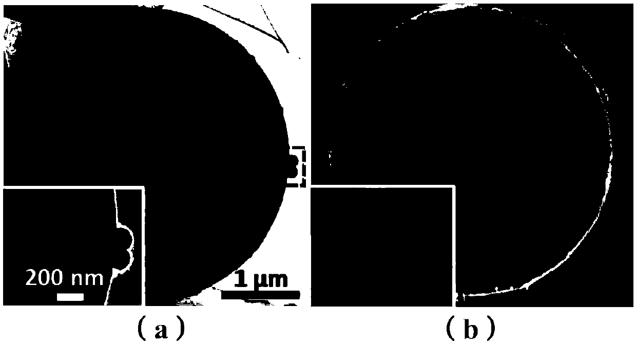
Embodiment 3
[0134] This example is used to illustrate the preparation and morphology characterization of exosome-microsphere complexes.
[0135] 1. Quantification of exosome protein
[0136] Take 20 μL of exosomes extracted according to the method of Example 1 in a centrifuge tube, add 20 μL of protein lysate (containing protease inhibitors), mix well, and lyse on ice for 1 hour, using the BCA protein quantification kit produced by Solebol Company The protein content of exosomes was quantified.
[0137] 2. Preparation of exosome-microsphere complexes
[0138] 1) According to the results of protein quantification, take exosomes with a protein content of 10 μg, mix them well with 2.5uL microspheres, incubate at room temperature for 15min, add PBS buffer to a total volume of 250ul, and incubate overnight at 4°C.
[0139] 2) Add 28 μL of glycine with a concentration of 1M to the above mixture, so that the final concentration of glycine is 100 mM, and react at room temperature for 30 minutes...
PUM
| Property | Measurement | Unit |
|---|---|---|
| Size distribution | aaaaa | aaaaa |
| Size | aaaaa | aaaaa |
Abstract
Description
Claims
Application Information