Preparation of biomimetic skeletal muscle composite tissue by multi-channel extrusion 3D bioprinting

A bioprinting, composite tissue technology, used in tissue regeneration, animal cells, prostheses, etc., can solve problems such as inability to combine, provide bone lesions, and muscles have no function.

Active Publication Date: 2021-08-20
福建省安悦莱生物科技有限公司
View PDF0 Cites 0 Cited by
  • Summary
  • Abstract
  • Description
  • Claims
  • Application Information

AI Technical Summary

Problems solved by technology

However autografts are limited by the amount of bone and may cause lesions at the donor bone site during reconstructive surgery
Although a flap involving bone and muscle can be transferred, the muscle is nonfunctional and cannot be combined with the supporting musculature

Method used


Image

  • Preparation of biomimetic skeletal muscle composite tissue by multi-channel extrusion 3D bioprinting
  • Preparation of biomimetic skeletal muscle composite tissue by multi-channel extrusion 3D bioprinting
  • Preparation of biomimetic skeletal muscle composite tissue by multi-channel extrusion 3D bioprinting

Examples


Embodiment 1

[0066] A bionic skeletal muscle composite tissue prepared by multi-channel extrusion 3D bioprinting, prepared by the following preparation method:

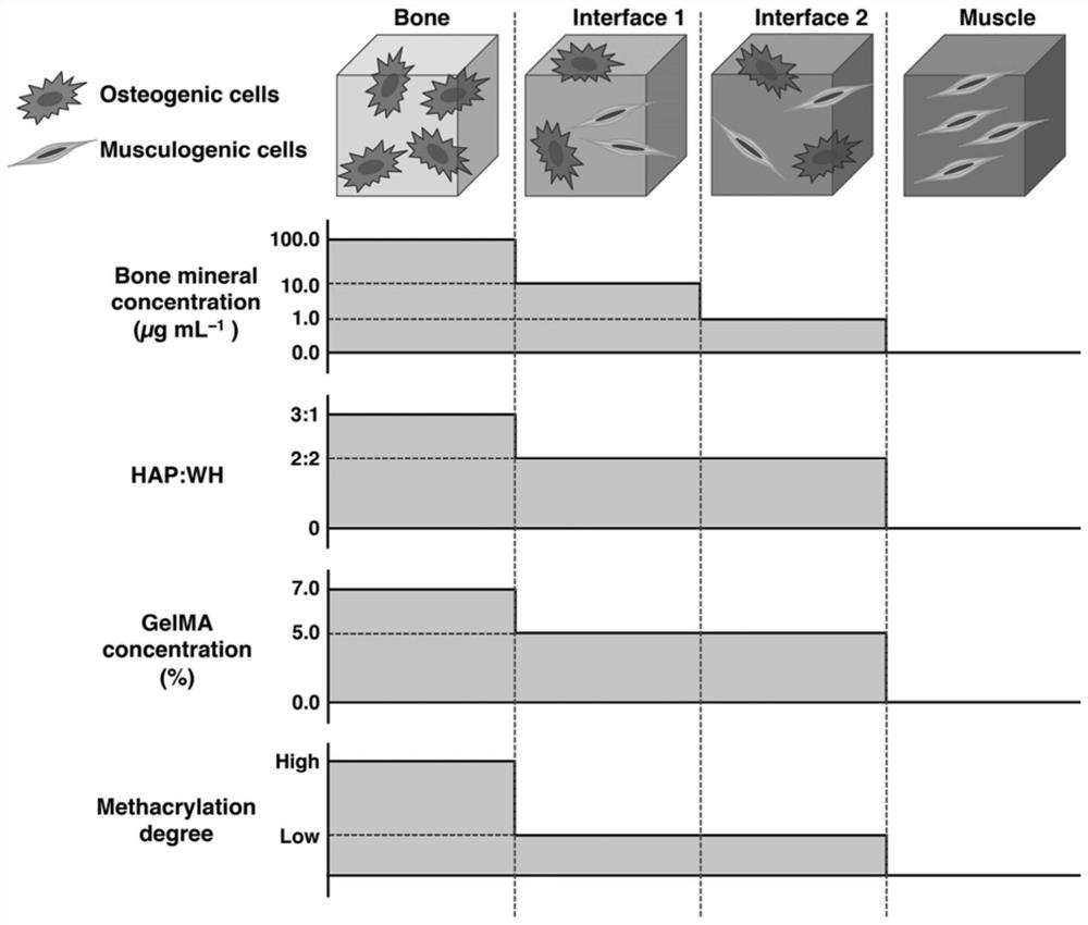
[0067] S1. By dropping MA into the gelatin aqueous solution, the substitution degree of MA in the bionic bone was 81.4%; the bionic periosteum was 19.7%; the bionic sarcolemma was 19.7%; the bionic muscle was 19.7%.

[0068] S2. The concentration of GelMA in the bionic bone is 7.0% ( W / V ), the concentration of SA is 0.5% ( W / V ) and a gelatin concentration of 3.0% ( W / V ); the concentration of GelMA in the bionic periosteum scaffold was 5.0% ( W / V ), the concentration of SA is 0.5% ( W / V ) and a gelatin concentration of 2.0% ( W / V ); the concentration of GelMA in the biomimetic sarcolemma scaffold was 5.0% ( W / V ), the concentration of SA is 0.5% ( W / V ) and a gelatin concentration of 2.0% ( W / V ); the concentration of GelMA in the bionic muscle scaffold was 5.0% ( W / V ), the concentration of SA is 0.5% ( W / V ) and a g...

Embodiment 2

[0075] A bionic skeletal muscle composite tissue prepared by multi-channel extrusion 3D bioprinting, prepared by the following preparation method:

[0076] S1. By dropping MA into the gelatin aqueous solution, the substitution degree of MA in the bionic bone is 90.0%; the bionic periosteum is 25.0%; the bionic sarcolemma is 25.0%; the bionic muscle is 25.0%.

[0077] S2. The concentration of GelMA in the bionic bone is 6.0% ( W / V ), the concentration of SA is 0.5% ( W / V ) and a gelatin concentration of 3.0% ( W / V ); the concentration of GelMA in the bionic periosteum scaffold was 4.0% ( W / V ), the concentration of SA is 0.5% ( W / V ) and a gelatin concentration of 2.0% ( W / V ); the concentration of GelMA in the biomimetic sarcolemma scaffold was 4.0% ( W / V ), the concentration of SA is 0.5% ( W / V ) and a gelatin concentration of 2.0% ( W / V ); the concentration of GelMA in the bionic muscle scaffold was 4.0% ( W / V), the concentration of SA is 0.5% ( W / V ) and gelatin ...

Embodiment 3

[0084] A biomimetic skeletal muscle composite tissue prepared by multi-channel extrusion 3D bioprinting is prepared by the following preparation method:

[0085] S1. By adding MA to gelatin aqueous solution, the substitution degree of MA in bionic bone is 81.4%; bionic periosteum is 19.7%; bionic sarcolemma is 19.7%; bionic muscle is 19.7%.

[0086] S2, the concentration of GelMA in the bionic bone is 7.0% ( W / V ), the concentration of SA is 0.5% ( W / V ) and gelatin at a concentration of 4.0% ( W / V ); the concentration of GelMA in the biomimetic periosteal scaffold was 5.0% ( W / V ), the concentration of SA is 0.5% ( W / V ) and gelatin at a concentration of 3.0% ( W / V ); the concentration of GelMA in the bionic sarcolemma scaffold was 5.0% ( W / V ), the concentration of SA is 0.5% ( W / V ) and gelatin at a concentration of 3.0% ( W / V ); the concentration of GelMA in the bionic muscle scaffold was 5.0% ( W / V ), the concentration of SA is 0.5% ( W / V ) and gelatin at a con...


PUM


Abstract

The invention discloses a bionic skeletal muscle composite tissue prepared by multi-channel extrusion 3D bioprinting. The preparation method comprises the following steps: preparing bone scaffold bionic bioink, periosteum bionic bioink, myofibrillar membrane bionic bioink, and muscle bionic bioink; Mix MSCs and C2C12 with the corresponding bionic bio-ink; use a multi-channel extrusion 3D bioprinter to print and form a four-layer composite tissue engineering scaffold of bionic bone, bionic periosteum, bionic sarcolemma, and bionic muscle. The bionic skeletal muscle composite tissue prepared by multi-channel extrusion 3D bioprinting of the present invention can minimize fibrosis during the recovery of traumatic skeletal muscle injury; the bionic skeletal muscle composite tissue prepared by multi-channel extrusion 3D bioprinting can simultaneously replace The structure and function of bone and skeletal muscle, supporting the proliferation and differentiation of myoblasts and osteoblasts; and using 3D bioprinting technology to make implants easy to customize to adapt to any defect shape.

Description

technical field [0001] The invention relates to the technical field of biomaterials, in particular to a bionic skeletal muscle composite tissue prepared by multi-channel extrusion 3D bioprinting. Background technique [0002] Skeletal muscle is one of the largest and most important organs in the human body, accounting for 45% of body weight (Choi, J. S. et al. Journal of Controlled Release. 2016, 222:107-115). More than one in two of these are affected by skeletal muscle injuries. Severe traumatic muscle injuries from motor vehicle accidents, crush injuries, and explosions are all causes of severe disability and lead to severe pain and lengthy recovery periods, adding to the economic burden of the patient (Yelin, E., et al . Seminars in Arthritis and Rheumatism. 46(3):259-260 ). Muscle function depends on proper insertion points on stable bone, so wounds where both bone and muscle are damaged heal particularly poorly. Although recently in bone — Progress has been made in...

Claims


Application Information

Patent Timeline
Patent Type & Authority Patents(China)
IPC IPC(8): C12N5/07A61L27/40A61L27/22A61L27/20A61L27/12A61L27/38A61L27/50B33Y10/00B33Y70/10B33Y80/00
CPCA61L27/12A61L27/20A61L27/222A61L27/3821A61L27/3826A61L27/3873A61L27/50A61L2430/30B33Y10/00B33Y70/00B33Y80/00C08L5/04
Inventor 张进黄恒童冬梅刘晓晨李飞翰
Owner 福建省安悦莱生物科技有限公司
Why Eureka
Try Eureka