
Fundus image-oriented microaneurysm detection method

A fundus image and detection method technology, applied in image enhancement, image analysis, image data processing, etc., can solve the problem of not taking into account the lesion information closely related to diagnosis, not taking into account the different importance of different layers of features, ignoring the target and surrounding Environmental correlation and other issues to achieve the effect of improving detection efficiency and screening out false detections

Inactive Publication Date: 2020-02-07
UNIV OF ELECTRONICS SCI & TECH OF CHINA +1
View PDF5 Cites 28 Cited by
  • Summary
  • Abstract
  • Description
  • Claims
  • Application Information

AI Technical Summary

Problems solved by technology

[0006] There are still deficiencies in the above-mentioned feature pyramid schemes. When performing feature fusion, the importance of different layers of features is not considered, and most target detection schemes mainly focus on the features of the target itself, thus ignoring the correlation between the target and the surrounding environment. The convolutional neural network is regarded as a black box, which does not take into account the lesion information closely related to the diagnosis

Method used


Image

  • Fundus image-oriented microaneurysm detection method
  • Fundus image-oriented microaneurysm detection method
  • Fundus image-oriented microaneurysm detection method

Examples


Embodiment Construction

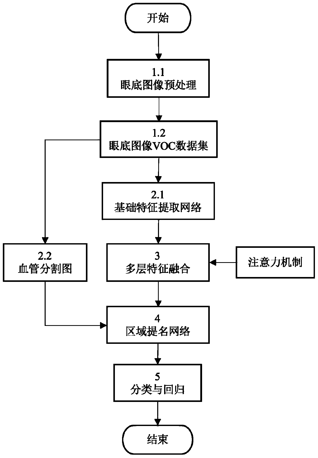
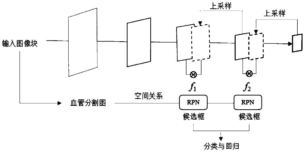
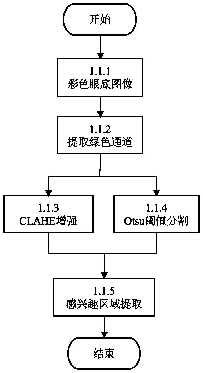
[0053] The purpose of the present invention is to address the shortcomings of micro-aneurysm detection in the prior art, and propose a micro-aneurysm detection method for fundus images, so as to effectively detect micro-aneurysms in fundus images and realize automatic detection At the same time, it can better assist doctors in making a diagnosis. Its core idea is: through the production of a series of fundus image preprocessing and detection data sets, the contrast of fundus images and the characteristics of microaneurysms are enhanced; The mechanism further enhances features useful for microaneurysm detection and suppresses noise. At the same time, the present invention also combines the positional relationship between microaneurysms and blood vessels to achieve the purpose of removing some false detections when selecting microaneurysm candidate frames. The present invention can automatically detect microaneurysms in fundus images based on deep learning, and uses the human v...


PUM


Abstract

The invention relates to a medical image processing technology, and provides a fundus image-oriented microaneurysm detection method for overcoming the defects of detection of a microaneurysm which isa micro target in the prior art, so as to effectively detect the microaneurysm in a fundus image, and better assist a doctor in diagnosis while realizing automatic detection. The method comprises: preprocessing the fundus image, so that the features of a small target are enhanced, and making and training a data set on a built basic feature extraction network; during detection, extracting image basic features from the input image, and performing blood vessel segmentation on the input image by utilizing a segmentation model to obtain a feature map and a segmentation map for subsequent processing; then integrating an attention mechanism into a feature fusion process to obtain a fused convolution feature layer; inputting the fused convolution feature layer into a candidate region generation network, and obtaining a candidate region by considering the position relationship between the target and the blood vessel; and finally, further classifying and regressing the candidate regions to obtain a detection result.

Description

technical field [0001] The invention relates to a medical image processing technology, in particular to a microaneurysm detection method for fundus images. Background technique [0002] Diabetic retinopathy (Diabetic Retinopathy, DR) is one of the most serious complications of diabetes, and it is also the main cause of blindness in the world. Microaneurysms (MAs) are the earliest detectable tiny lesions in the early stage of DR lesions, and appear as small dark red dots in retinal fundus images. The traditional screening of DR lesions mainly relies on the diagnosis of retinal fundus images by ophthalmologists, which not only requires high professional skills of doctors, but also the diagnosis process is time-consuming and labor-intensive. As the incidence of diabetes continues to rise, medical resources are increasingly scarce. Therefore, automatic detection of microaneurysms is of great significance for DR screening and assisting doctors in diagnosis. [0003] In the pri...

Claims


Application Information

Patent Timeline
Patent Type & Authority Applications(China)
IPC IPC(8): G06T5/40G06T7/00G06T7/11G06T7/136G06T7/90G06K9/32G06K9/62G06N3/04G06N3/08
CPCG06T7/11G06T7/136G06T7/0012G06T7/90G06T5/40G06N3/08G06T2207/30041G06V10/25G06N3/045G06F18/24
Inventor 詹开明罗光春连春华吴钒田玲段贵多李英董代宇
Owner UNIV OF ELECTRONICS SCI & TECH OF CHINA
Learn More