Recombinant anti-PD-L1 monoclonal antibody

A monoclonal antibody, PD-L1 technology, applied in the field of biomedicine, can solve the problem of no obvious response and achieve high affinity effect

Pending Publication Date: 2020-03-13
SHANGHAI ZHANGJIANG BIOTECH
View PDF6 Cites 6 Cited by
  • Summary
  • Abstract
  • Description
  • Claims
  • Application Information

AI Technical Summary

Problems solved by technology

Therefore, it can be seen that more than half of the patients have no obvious response to anti-PD-L1 treatment (this may be related to factors such as individual genetic diversity, tumor type

Method used


Image

  • Recombinant anti-PD-L1 monoclonal antibody
  • Recombinant anti-PD-L1 monoclonal antibody
  • Recombinant anti-PD-L1 monoclonal antibody

Examples


Embodiment 1

[0060] Embodiment 1, construction and screening of hybridoma cells

[0061] 1. Immunized animals

[0062] Use the recombinantly expressed PD-L1-Fc fusion protein (fused from the extracellular region of human PD-L1 to the Fc segment of the antibody, the amino acid sequence is the sequence numbered Q9NZQ7-1 on the UniProt website) with a dose of 100-500 μg / mL intraperitoneally Immunize Balb / c mice for 3 times. 3-7 days before cell fusion, boost immunization with PD-L1-Fc fusion protein 50-100 μg / mL tail vein.

[0063] 2. Cell Fusion

[0064] The mouse myeloma cell line NS-1 was fused with the splenocytes of Balb / c mice after immunization, and then placed in a 96 empty cell culture plate, screened with a complete medium containing HAT, half of the medium was changed in 3-5 days, 2 Colony formation can be seen in about a week.

[0065] 3. Identification of culture supernatant

[0066] The hybridoma cell culture supernatant was detected by ELISA technique. Coat multi-well pla...

Embodiment 2

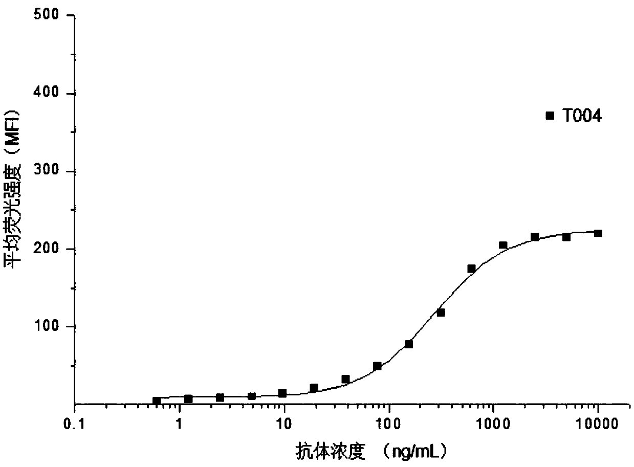
[0071] Example 2. Flow cytometry detection of the binding ability of T0004 antibody to PD-L1 on the cell membrane surface

[0072] The expression vector plasmid containing the full-length coding sequence of human PD-L1 was stably transfected into CHO-K1 host cells, and CHO-K1 engineered cells stably expressing PD-L1 on the membrane surface were obtained by pressurized screening (denoted as CHO-K1 / hmPD- L1). T0004 antibody was diluted to 15 concentrations starting from 10 μg / mL, mixed with CHO-K1 / hmPD-L1 cells in the logarithmic growth phase, and kept on ice for 45 min to make it adhere to the surface of CHO-K1 / hmPD-L1 cells PD-L1 binding. Wash 2 times with PBS containing 1% fetal bovine serum, add FITC fluorescently labeled rabbit anti-mouse IgG (H+L) secondary antibody, ice bath for 45min, wash 2 times with PBS containing 1% fetal bovine serum. The pellet was resuspended in PBS containing 1% fetal bovine serum, and the fluorescence intensity was analyzed by flow cytometry. ...

Embodiment 3

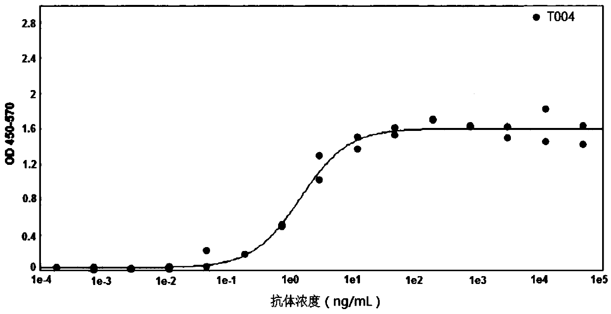
[0074] Example 3. Enzyme-linked immunoassay detection of binding ability of T0004 antibody to soluble PD-L1

[0075] Dilute the human PD-L1-Fc fusion protein to 10 μg / mL with coating solution, add 100 μL / well to the microtiter plate, and coat at 37°C for 1-2 hours. Discard the coating solution, add blocking solution, 350 μL / well, block overnight at 2-8°C, and wash 7 times with a plate washer. Dilute the T0004 antibody with PBS, starting from 50000ng / mL, dilute 15 concentrations at a 4-fold ratio, 100μL / well, react at 37°C for 1-2 hours, discard the liquid in the microplate plate, and wash 7 times with a plate washer. Add horseradish peroxidase (HRP)-labeled goat anti-mouse IgG (H+L) diluted 1:10000, 100 μL / well, react at 37°C for 1 hour, discard the liquid in the microtiter plate, and wash 7 times with a plate washer . Add TMB substrate solution to the ELISA plate, 50 μL / well, develop color in the dark for 1-10 minutes, add 50 μL / well stop solution, and mix quickly. Read OD...


PUM


Abstract

The invention discloses a recombinant anti-PD-L1 monoclonal antibody, relates to the technical field of biological medicines, and is used for providing an effective treatment medicine for patients with advanced or metastatic cancers, in particular to the patients with ineffective or drug-resistant treatment by an existing anti-PD-L1 medicine. The complementarity determining region of the antibodyhas sequences shown in SEQ ID NO: 1 to SEQ ID NO: 6. Compared with an existing anti-PD-L1 drug, the antibody has a unique binding epitope, and is better in affinity to human PD-L1 and better in tumorinhibition effect.

Description

technical field [0001] The invention relates to the technical field of biomedicine, and more specifically, the invention discloses a monoclonal antibody suitable for the treatment or detection of cancer and immune diseases. Background technique [0002] PD-L1 (Programmed cell death 1 ligand 1, programmed cell death ligand 1) is also known as surface antigen differentiation cluster 274 (cluster of differentiation 274, CD274) or B7 homolog 1 (B7 homolog 1, B7-H1) , is a transmembrane protein with a size of 40kDa, one of the two ligands of PD-1 (Programmed Cell Death Protein 1), highly expressed in the heart, skeletal muscle, placenta, and lung, and in the thymus, spleen, kidney, and liver Low expression, also expressed in activated T cells, B cells, dendritic cells and other immune cells, and widely expressed on a variety of tumor cells. [0003] The combination of PD-L1 and PD-1 can transmit immunosuppressive signals and reduce the proliferation of T cells. This "immune chec...

Claims


Application Information

Patent Timeline
IPC IPC(8): C07K16/28A61K39/395A61P35/00A61P37/02G01N33/68G01N33/574
CPCC07K16/2827A61P35/00A61P37/02G01N33/6893G01N33/574C07K2317/24C07K2317/732C07K2317/734C07K2317/92C07K2317/94A61K2039/505G01N2800/24
Inventor 聂丽刘广洛
Owner SHANGHAI ZHANGJIANG BIOTECH
Why Eureka
Try Eureka