
Preparation method of bacteriophage transmission electron microscope specimen

A technique of transmission electron microscopy and bacteriophage, which is applied in material analysis using radiation diffraction, material analysis using radiation, and material analysis using wave/particle radiation, etc. The effect of saving manpower, material resources and cost, and high throughput

Inactive Publication Date: 2020-03-24
云南省地方病防治所
View PDF6 Cites 0 Cited by
  • Summary
  • Abstract
  • Description
  • Claims
  • Application Information

AI Technical Summary

Problems solved by technology

In the process of phage research, identifying the morphology of the phage is a prerequisite. At present, there are many methods for morphological identification of phage reported in domestic and foreign research, mainly focusing on the application of different types of electron microscopy. There are relatively few methods for preparing phage samples, and Most of the time is too long, and the amount of specimens and reagents is large. Therefore, it is very necessary to find a high-quality method for preparing phage transmission electron microscope specimens.

Method used


Image

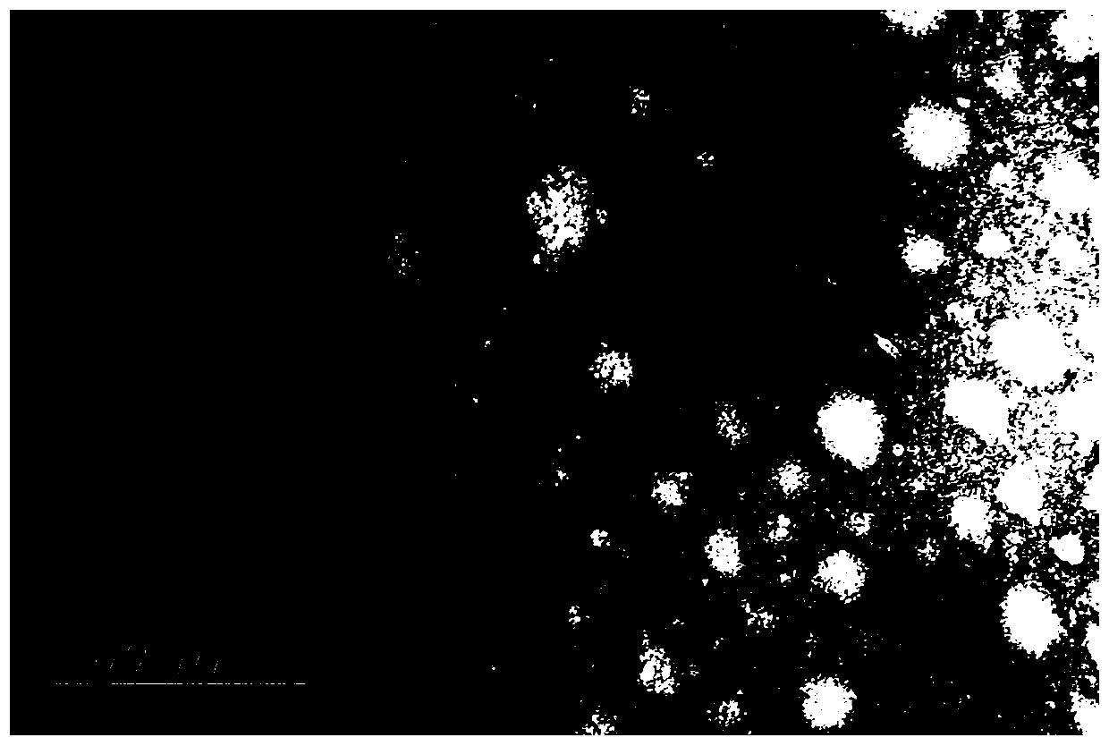
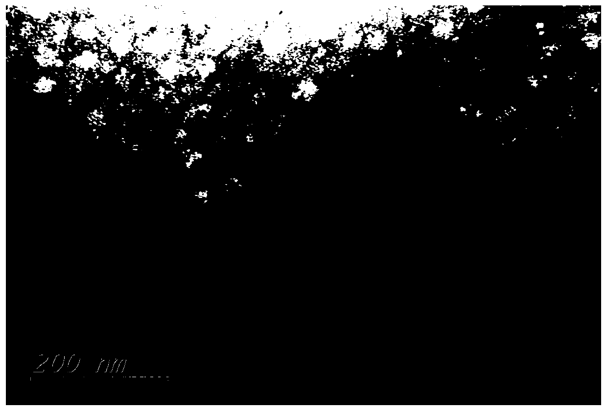
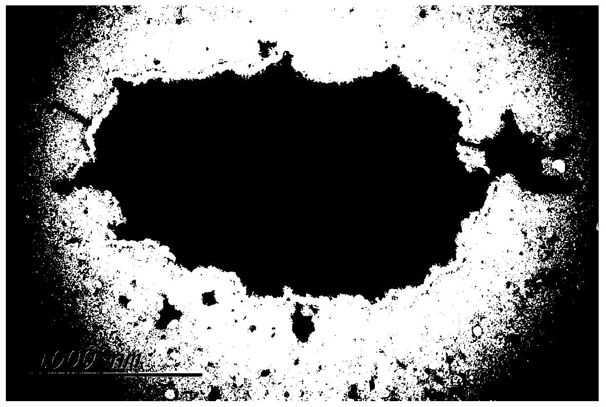
  • Preparation method of bacteriophage transmission electron microscope specimen
  • Preparation method of bacteriophage transmission electron microscope specimen
  • Preparation method of bacteriophage transmission electron microscope specimen

Examples


Embodiment 1

[0024] A method for preparing a bacteriophage transmission electron microscope specimen is further illustrated by taking plague phage as an example, and specifically includes the following steps:

[0025] (1) After the purified wild plague phage stored at 4°C was rejuvenated at 28°C, 220rpm, and 24h, the titer was measured, and the titer reached 10 8 Above, proceed to the next step of specimen preparation.

[0026] (2) Spread the host bacteria EV76 (plague vaccine strain) on a double-layer plate, incubate at 28°C for 1.5 hours, add 15 μL of the above-mentioned phage dropwise, and after it is completely absorbed, continue to culture at 28°C to make the phage plaques clear.

[0027] (3) Use a 10 μL pipette tip to pick out the upper layer of agar containing phage plaques and put it into a 1.5ml centrifuge tube with 200 μL of normal saline (or SM solution) to mash, place at 4°C for 2 hours, 5000 rpm, 2 minutes, and absorb the supernatant.

[0028] (4) Prepare a piece of parafilm ...

Embodiment 2

[0034] A method for preparing a bacteriophage transmission electron microscope specimen is further illustrated by taking Escherichia coli ATCC8739 as an example, and specifically includes the following steps:

[0035] (1) Measure the titer of the purified coliphage stored at 4°C after rejuvenation at 37°C, 220rpm, and 24h, and the titer needs to reach 10 8 Above, the next step of specimen preparation can be carried out.

[0036] (2) Spread the host bacteria (Escherichia coli ATCC8739) on a double-layer plate, incubate at 37°C for 3 hours, add 15 μL of the above-mentioned phages dropwise, and continue to culture at 37°C until the phage plaques become clear.

[0037] (3) Use a 10 μL pipette tip to pick out the upper layer of agar containing phage plaques and put it into a 1.5ml centrifuge tube with 200 μL of normal saline (or SM solution) to mash, place at 4°C for 2 hours, 5000 rpm, 2 minutes, and absorb the supernatant.

[0038] (4) Prepare a piece of parafilm or clean film gl...

Embodiment 3

[0044] A method for preparing a bacteriophage transmission electron microscope specimen is further illustrated by taking Staphylococcus aureus ATCC25923 as an example, specifically comprising the following steps:

[0045] (1) Measure the titer of the purified Staphylococcus aureus phage stored at 4°C after rejuvenation at 37°C, 220rpm, and 24h, and the titer needs to reach 10 8 Above, the next step of specimen preparation can be carried out.

[0046] (2) Spread the host bacteria (Staphylococcus aureus ATCC25923) on a double-layer plate, incubate at 37°C for about 1 to 3 hours, add 15 μL of the above-mentioned phage dropwise, and continue to culture at 37°C until the plaque becomes clear after it is completely absorbed.

[0047] (3) Use a 10 μL pipette tip to pick out the upper layer of agar containing phage plaques and put it into a 1.5ml centrifuge tube with 200 μL of normal saline (or SM solution) to mash, place at 4°C for 2 hours, 5000 rpm, 2 minutes, and absorb the supernata...


PUM


Abstract

The invention discloses a preparation method of a bacteriophage transmission electron microscope specimen, and belongs to the technical field of bacteriophage specimen preparation. The method disclosed by the invention comprises the following steps: rejuvenating purified bacteriophage to make the titer reach 106 or above; paving host bacteria on a double-layer flat plate for culturing until the bacteriophage plaque is transparent; taking out upper agar containing bacteriophage plaque, carrying out centrifugal treatment, sucking supernatant, taking the front surface of the copper net, upwards putting the copper net into bacteriophage liquid drops, dyeing, taking out the copper net after dyeing, sucking residual dye liquor, putting the copper net back to the copper net box, and marking the position to obtain the bacteriophage transmission electron microscope specimen. 10 parts of samples can be prepared in one week in a traditional method, 100 parts of samples can be prepared in two daysin the method, the flux is high, meanwhile, the action condition of the bacteriophage and a host can be observed, the bacteriophage can be used immediately after being expanded, and the form of the bacteriophage in a fresh state can be ensured.

Description

technical field [0001] The invention discloses a preparation method of a bacteriophage transmission electron microscope specimen, belonging to the technical field of phage specimen preparation. Background technique [0002] Phages were discovered independently by Twort and D. Herelle in 1907 and 1909 respectively. Phage (bacteriophage, phage) is a general term for viruses that infect microorganisms such as bacteria, fungi, actinomycetes, or spirochetes. Some of them can cause lysis of host bacteria, so they are called phages. Bacteriophages are widely distributed, and wherever there are bacteria, corresponding phages may exist. Phages of intestinal bacteria are often contained in human and animal excrement or polluted well water and river water. In the soil, phages of soil bacteria can be found; phages have strict host specificity and only live in susceptible hosts, so phages can be used for epidemiological identification and typing of bacteria to trace the source of infec...

Claims


Application Information

Patent Timeline
Patent Type & Authority Applications(China)
IPC IPC(8): G01N23/04G01N23/20G01N23/2202
CPCG01N23/04G01N23/20G01N23/2202
Inventor 钟佑宏李伟王鹏黄英张海鹏段存娟杨丰义
Owner 云南省地方病防治所
Learn More