Cell fluorescence in situ hybridization method based on microfluidic chip and application thereof

A fluorescence in situ hybridization and microfluidic chip technology, applied in the field of microfluidics, can solve the problems of difficult automatic operation, large reagent consumption, complicated operation steps, etc., achieve good fluorescence in situ hybridization effect and save reagent consumption , Analyzing the effect of high detection sensitivity

Pending Publication Date: 2020-04-03
SHENZHEN INST OF ADVANCED TECH CHINESE ACAD OF SCI
View PDF9 Cites 1 Cited by
  • Summary
  • Abstract
  • Description
  • Claims
  • Application Information

AI Technical Summary

Problems solved by technology

In the fluorescence in situ hybridization method, the reagent consumption of the fluorescence in situ hybridization method is large, the operation steps are complicated, it is not easy to realize automatic operation, and the fluorescence background is high

Method used


Image

  • Cell fluorescence in situ hybridization method based on microfluidic chip and application thereof
  • Cell fluorescence in situ hybridization method based on microfluidic chip and application thereof
  • Cell fluorescence in situ hybridization method based on microfluidic chip and application thereof

Examples


preparation example Construction

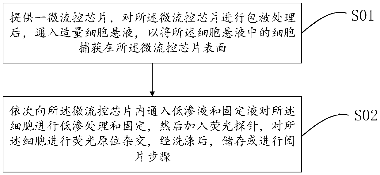
[0038] Optionally, the preparation method of the microfluidic chip includes:

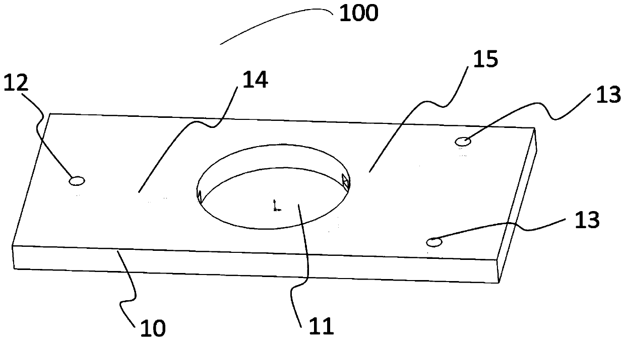
[0039] A patterned hard template and a flow channel layer substrate are provided, combined with imprinting technology, the flow channel layer substrate is imprinted, punched and cleaned to form cell capture holes on the flow channel layer substrate, at least one further The pattern structure of the sample hole, at least one sample outlet hole, the first flow channel and the second flow channel;

[0040]A sealing layer substrate is provided, and a microfluidic chip is obtained by aligning and bonding the sealing layer substrate and the embossed, punched and cleaned flow channel layer substrate, and heat-pressing and sealing them.

[0041] Optionally, the materials of the channel layer substrate and the sealing layer substrate include polydimethylsiloxane (PDMS), cycloolefin copolymer, polystyrene, polymethacrylate, polyterephthalic acid One or more of glycol ester, polytetrafluoroethylene, polypropi...

Embodiment 1

[0070] A cell fluorescence in situ hybridization method based on a microfluidic chip, comprising:

[0071] A microfluidic chip is provided, and the microfluidic chip is coated with 0.1% polylysine; the cultured A549 cells (human non-small cell lung cancer cells) are digested, resuspended, diluted to a certain concentration and passed through into the microfluidic chip;

[0072] After passing through the cells, the hypotonic solution (0.4% potassium chloride and 0.8% sodium citrate mixed at a volume ratio of 1:1) was passed into the microfluidic chip in turn to perform hypotonic treatment on the A549 cells, and after 10 minutes, continue to pass through A549 cells were fixed in a fixative solution (mixed with absolute ethanol and glacial acetic acid at a volume ratio of 3:1) for 8 min; then 70%, 90% and 100% ethanol were sequentially passed into the microfluidic chip for 1 min; after that, CEP8 -The FITC probe solution was mixed and passed into the microfluidic chip, denatured...

Embodiment 2

[0074] A cell fluorescence in situ hybridization method based on a microfluidic chip, comprising:

[0075]Provide a microfluidic chip, use 0.1% polylysine to coat the microfluidic chip; pass the fetal nucleated red blood cell sample cells into the microfluidic chip;

[0076] After passing through the cells, the hypotonic solution (0.4% potassium chloride and 0.8% sodium citrate mixed at a volume ratio of 1:1) was sequentially passed into the microfluidic chip to perform hypotonic treatment on the fetal nucleated red blood cells. After 10 minutes, Continue to pass through the fixation solution (mixture of absolute ethanol and glacial acetic acid at a volume ratio of 3:1) to fix the fetal nucleated red blood cells for 8 minutes; then use 70%, 90% and 100% ethanol to pass through the microfluidic chip for 1 minute. ; Then mix the CEP8-FITC probe solution and pass it into the microfluidic chip, denature at 75°C for 12 minutes; hybridize overnight at 42°C; after the hybridization i...


PUM


Abstract

Embodiment of the invention provides a cell fluorescence in situ hybridization method based on a microfluidic chip. The method includes the following steps: providing a microfluidic chip; passing an appropriate amount of cell suspension into the microfluidic chip after the microfluidic chip being coated to capture cells in the cell suspension on the surface of the microfluidic chip; passing a hypotonic solution and a fixing solution into the microfluidic chip sequentially to perform hypotonic treatment and fixation on the cells, and then adding fluorescent probes to perform fluorescence in situ hybridization on the cells; conducting storing or reading step after washing. The cell fluorescence in situ hybridization method has the advantages of simple operation steps, low reagent consumption, low cost and low fluorescence background. The invention also provides application of the cell fluorescence in situ hybridization method based on the microfluidic chip.

Description

technical field [0001] The invention relates to the field of microfluidic technology, in particular to a microfluidic chip-based cell fluorescence in situ hybridization method and its application. Background technique [0002] Fluorescence in situ hybridization (FISH) is a new technology combining non-radioactive molecular biology and cytogenetics developed on the basis of radioactive in situ hybridization in the late 1980s. It is replaced by fluorescent markers. A new in situ hybridization method formed by isotope labeling. [0003] At present, fluorescence in situ hybridization of cells is mainly carried out on glass slides, and the cells are fixed on the glass slides before performing operations such as hybridization, washing, and slide reading. In the fluorescence in situ hybridization method, the reagent consumption of the fluorescence in situ hybridization method is large, the operation steps are complicated, it is difficult to realize automatic operation, and the flu...

Claims


Application Information

Patent Timeline
Patent Type & Authority Applications(China)
IPC IPC(8): C12Q1/6841
CPCC12Q1/6841C12Q2563/107C12Q2565/629C12Q2565/631
Inventor 黄玉清陈艳
Owner SHENZHEN INST OF ADVANCED TECH CHINESE ACAD OF SCI
Why Eureka
Try Eureka