Supercharge Your Innovation With Domain-Expert AI Agents!

Quality control method of medical image, computer equipment and readable storage medium

A medical image and quality control method technology, applied in the field of image processing, can solve the problems of low accuracy and efficiency of quality control results

Pending Publication Date: 2020-04-28
SHANGHAI UNITED IMAGING INTELLIGENT MEDICAL TECH CO LTD
View PDF9 Cites 8 Cited by
  • Summary
  • Abstract
  • Description
  • Claims
  • Application Information

AI Technical Summary

Problems solved by technology

[0005] Based on this, it is necessary to provide a medical image quality control method, computer equipment and readable storage medium for the problem of low accuracy and efficiency of quality control results in traditional technologies

Method used


Image

  • Quality control method of medical image, computer equipment and readable storage medium
  • Quality control method of medical image, computer equipment and readable storage medium
  • Quality control method of medical image, computer equipment and readable storage medium

Examples


Embodiment Construction

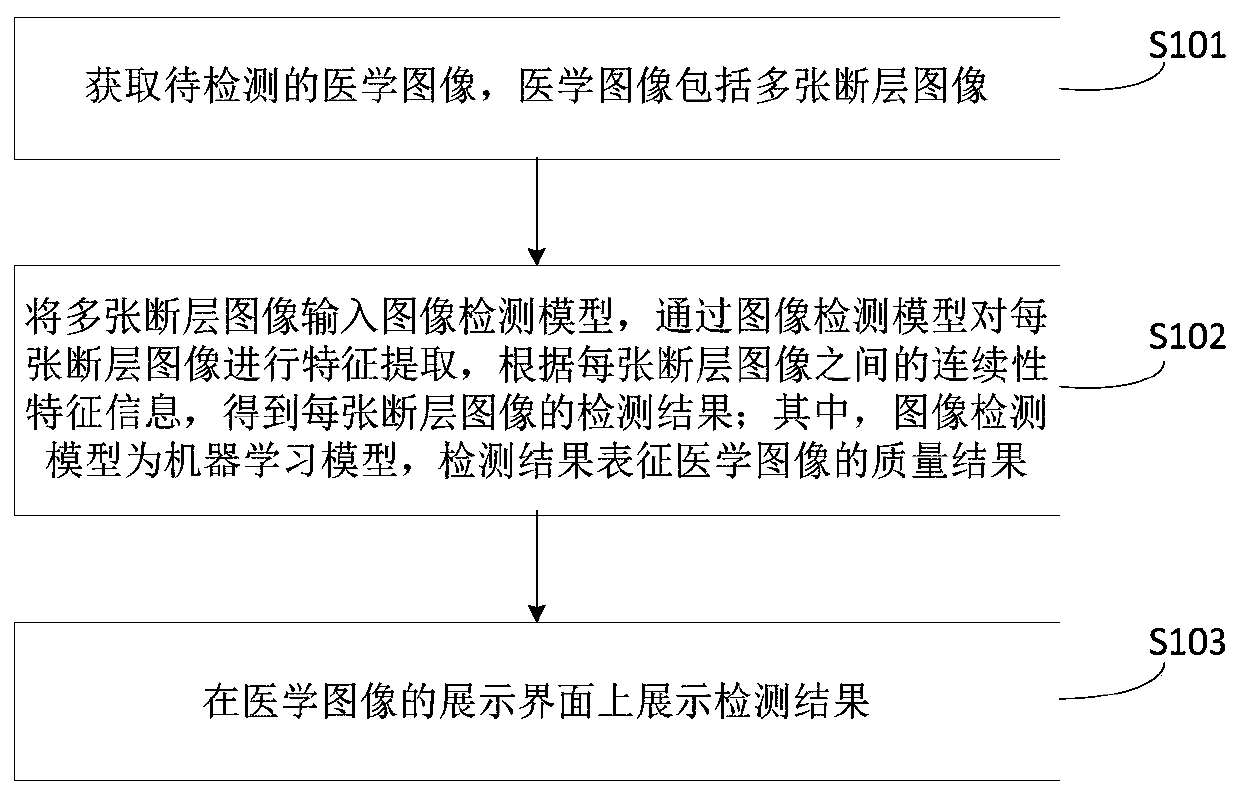
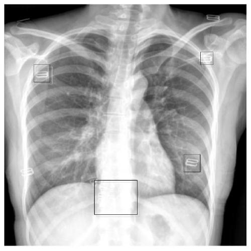
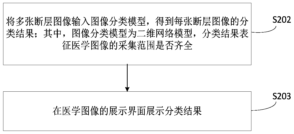
[0054]The quality control method of the medical image provided by the embodiment of the present application can be applied to the quality control process of the medical image, and the medical image can be direct digital flat panel X-ray imaging (Digital Radiography, DR), electronic computer tomography , CT), nuclear magnetic resonance imaging (Nuclear Magnetic Resonance Imaging, MRI), positron emission computed tomography (Positron Emission Computed Tomography, PET), etc. Taking CT as an example, contrast-enhanced scanning is usually used to determine whether there is a mass or enlarged lymph node in the mediastinum and hilum, whether there is stenosis or obstruction in the bronchi, and to detect primary and metastatic mediastinal tumors, lymph node tuberculosis, and central lung cancer. With greater help, interstitial and solid lesions in the lungs can also be displayed better. However, due to the current medical level is still uneven, the CT imaging technology of lower-level...


PUM


Abstract

The invention relates to a medical image quality control method, computer equipment and a readable storage medium. The method comprises the following steps: obtaining a medical image to be detected, wherein the medical image comprises a plurality of tomographic images; inputting the plurality of tomographic images into an image detection model, performing feature extraction on each tomographic image through the image detection model, and obtaining a detection result of each tomographic image according to continuity feature information between the tomographic images; wherein the image detectionmodel is a machine learning model, and the detection result represents the quality result of the medical image; and displaying the detection result on a display interface of the medical image. According to the method, after the detection result is obtained, the detection result is displayed to the corresponding technician in time, the technician can know the problems existing during shooting andcorrect the problems in time, and therefore the quality of the obtained medical image can be greatly improved. Furthermore, the process of obtaining the medical image detection result is executed by the image detection model, manual participation is not needed, and the accuracy and efficiency of quality control are greatly improved.

Description

technical field [0001] The present application relates to the technical field of image processing, in particular to a medical image quality control method, computer equipment and a readable storage medium. Background technique [0002] With the continuous development of medical technology, the continuous improvement of social medical system and the improvement of people's living standards, computerized tomography (CT) image diagnosis technology has become an important equipment for human disease diagnosis. X-ray beams, γ-rays, ultrasonic waves, etc., together with highly sensitive detectors, perform cross-sectional scanning around a certain part of the human body one by one. It has the characteristics of fast scanning time and clear images. However, due to the uneven level of medical care in China, the quality of CT images obtained is also uneven, and images with poor quality will directly affect the follow-up doctor's diagnosis and patient treatment. [0003] The tradition...

Claims


Application Information

Patent Timeline
IPC IPC(8): G06T7/00G06K9/62
CPCG06T7/0012G06T2207/10072G06T2207/10081G06T2207/20081G06T2207/20084G06T2207/30168G06F18/24Y02P90/30
Inventor 伍吉兵郑介志
Owner SHANGHAI UNITED IMAGING INTELLIGENT MEDICAL TECH CO LTD
Features
  • R&D
  • Intellectual Property
  • Life Sciences
  • Materials
  • Tech Scout
  • Unparalleled Data Quality
  • Higher Quality Content
  • 60% Fewer Hallucinations
Learn More