Preparation method of personalized customization type craniomaxillofacial bone surgery repair and reconstruction implant

A craniofacial and orthopedic surgery technology, applied in bone implants, joint implants, joint implants, etc., can solve the problems of difficult to repair structural defects, the strength of titanium mesh cannot meet the requirements, etc., to improve efficiency Effect

Inactive Publication Date: 2020-05-01
SHAANXI HENGTONG INTELLIGENT MACHINE
View PDF9 Cites 3 Cited by
  • Summary
  • Abstract
  • Description
  • Claims
  • Application Information

AI Technical Summary

Problems solved by technology

Although this method can make a more accurate restoration model, because the titanium mesh is pressed against the restoration model to make the restoration, it is difficult to make a fitting restoration for defects with complex structures. For

Method used


Image

  • Preparation method of personalized customization type craniomaxillofacial bone surgery repair and reconstruction implant
  • Preparation method of personalized customization type craniomaxillofacial bone surgery repair and reconstruction implant
  • Preparation method of personalized customization type craniomaxillofacial bone surgery repair and reconstruction implant

Examples


preparation example Construction

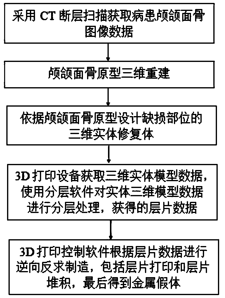
[0029] refer to figure 1 , 2 , 3, and 4, the present invention specifically discloses a method for preparing a personalized and customized craniofacial surgical repair and reconstruction implant, comprising the following steps:

[0030] Step 1: Obtain the three-dimensional data of the cranio-maxillofacial bone prototype: use X-ray film or CT scan to obtain the image data of the patient's cranio-maxillofacial bone, record the data into a CD-ROM file in standard DICOM format, and store it in the general medical image control system software A computer reads craniomaxillofacial bone image data from the storage medium;

[0031] Step 2: 3D reconstruction of the cranio-maxillofacial bone prototype, processing the tomographic image data of the cranio-maxillofacial bone in the CT image, including filtering, roughness reduction-grayscale image binarization-contour extraction-vectorized 3D modeling, using STL format to output the 3D model data of the craniofacial bone prototype;

[0...

Embodiment 1

[0042] Embodiment 1 (the design of the mandibular substitute of right half side defect Figure 5 )

[0043] Include the following steps:

[0044] Step 1: Obtain the three-dimensional data of the mandible prototype: use X-ray film or CT scan to obtain the tomographic image data of the patient's mandible, record the data in a standard DICOM format as a CD-ROM file, and store it in the computer of the general medical image control system software. The storage medium reads in the tomographic image data of the mandible;

[0045] Step 2: 3D reconstruction of the mandible prototype, preprocessing the tomographic image data of the mandible in the CT image, including image filtering, image smoothing and image denoising; binarizing the preprocessed grayscale image, the purpose of which is to Image segmentation is mainly the segmentation of bone tissue and soft tissue; after the above processing, the single-valued area of ​​the target contour can be obtained, and the outer contour value ...

Embodiment 2

[0056] Embodiment 2 (the typical structure of the mandibular substitute of left hemi defect Figure 6 )

[0057] Include the following steps:

[0058] Step 1: Obtain the three-dimensional data of the mandible prototype: use X-ray film or CT scan to obtain the tomographic image data of the patient's mandible, record the data in a standard DICOM format as a CD-ROM file, and store it in the computer of the general medical image control system software. The storage medium reads in the tomographic image data of the mandible;

[0059] Step 2: 3D reconstruction of the mandible prototype, preprocessing the tomographic image data of the mandible in the CT image, including image filtering, image smoothing and image denoising; binarizing the preprocessed grayscale image, the purpose of which is to Image segmentation is mainly the segmentation of bone tissue and soft tissue; after the above processing, the single-valued area of ​​the target contour can be obtained, and the outer contour...


PUM

PropertyMeasurementUnit
Thicknessaaaaaaaaaa
Thicknessaaaaaaaaaa

Abstract

The invention discloses a preparation method of a personalized customization type craniomaxillofacial bone surgery repair and reconstruction implant based on patient CT image design and repair body assembly. The preparation method is characterized in that after a computer storing medical image control system software and reverse engineering design software performs data segmentation on a tomography image, a patient head 3D prototype is reconstructed, a repair body for a coloboma part is designed according to the 3D prototype, 3D printing equipment acquires 3D entity model data, and hierarchy software is used to perform hierarchical processing on the 3D entity model data to obtain hierarchy data; and 3D printing control software performs reverse engineering manufacturing according to the hierarchy data, including hierarchy printing and hierarchy accumulation to obtain a metal prosthesis finally.

Description

technical field [0001] The present invention relates to a preparation method of an artificial prosthesis used to replace craniomaxillofacial bone defects, in particular to a preparation method of a personalized and customized craniomaxillofacial surgical repair and reconstruction implant, which adopts advanced manufacturing technology for personalization The processing of cranio-maxillofacial bone prosthesis is suitable for the repair of various bone defects and bone deformities caused by benign tumors, trauma and other reasons in any part of the cranio-maxillofacial region, and realizes the precise positioning of the repaired defect. Background technique [0002] Traditional repair methods mainly include prosthesis, implant prosthesis, surgical repair of skin flap and bone flap transplantation, and craniofacial bone traction technology. The above repair methods all have certain disadvantages in terms of postoperative shape and function, and cannot achieve shape repair and f...

Claims


Application Information

Patent Timeline
IPC IPC(8): A61F2/28A61F2/30G06T17/00
CPCA61F2/28A61F2/30942G06T17/00A61F2002/30948A61F2002/30952A61F2002/30985A61F2002/3097A61F2002/30971
Inventor 康柱郑力铭佘淮滨
Owner SHAANXI HENGTONG INTELLIGENT MACHINE
Why Eureka
Try Eureka