Application of skin tissue immune cell in situ detection kit for lupus erythematosus classification

A lupus erythematosus and immune cell technology, applied in the field of immune detection, can solve the problems of difficult flow analysis of single cell suspension cell number, inability to realize multiple labeling of tissue in situ markers, lack of spatial distribution information, etc.

Active Publication Date: 2020-06-26
THE SECOND XIANGYA HOSPITAL OF CENT SOUTH UNIV
View PDF9 Cites 0 Cited by
  • Summary
  • Abstract
  • Description
  • Claims
  • Application Information

AI Technical Summary

Problems solved by technology

For the in situ detection of immune cells in skin tissue, the traditional immunohistochemical single-staining or double-staining techniques have limitations, and cannot realize multiple labeling of tissue in situ markers, and cannot simultaneously observe and quantitatively detect various immune cells
Although methods such as flow cytometry and next-generation sequencing can identify cell types in skin tissue, the tissue must first be dissociated into a single-cell suspension before proceeding.
Clinically, human skin tissue samples are difficult to obtain, and the volume of the sample is generally small. The prepared single-cell suspension is often difficult to perform flow analysis due to the small number of cells, and is easy to be contaminated and degraded, making it difficult to complete sequencing.
Moreover, the results obtained by methods such as flow cytometry lack corresponding spatial distribution information, and cannot visually display its histomorphological data.
In addition, although some studies have shown that there are certain differences in certain immune cells between subtypes of lupus erythematosus, due to the limitations of traditional staining techniques, multiple labeling and quantitative detection cannot be achieved. Related application of diagnosis and classification of lupus erythematosus

Method used


Image

  • Application of skin tissue immune cell in situ detection kit for lupus erythematosus classification
  • Application of skin tissue immune cell in situ detection kit for lupus erythematosus classification
  • Application of skin tissue immune cell in situ detection kit for lupus erythematosus classification

Examples


Embodiment 1

[0083] Example 1: Preparation method of skin tissue in situ multi-label immunostaining kit

[0084] The results of multi-label immunostaining are captured through the processing of filters of four color channels (DAPI, FITC, Cy3, Cy5), and there will be some overlap between the emission spectra of different filters. The multi-spectral analysis software is based on the peak of the spectrum when identifying and splitting the spectrum. When multiple monoclonal antibodies are co-stained on a tissue section, the spectra corresponding to multiple antibodies will overlap to varying degrees. When an antibody develops too much color, its chromogenic spectrum may cover the spectrum of adjacent antibodies, resulting in cross-coloring between two or more antibodies. In order to avoid this situation, in the preparation of the skin tissue in situ multi-label immunostaining kit, the condition optimization of each key step is very important.

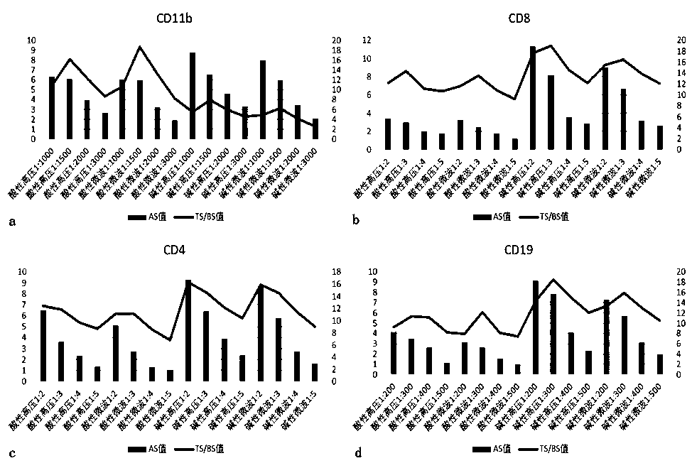
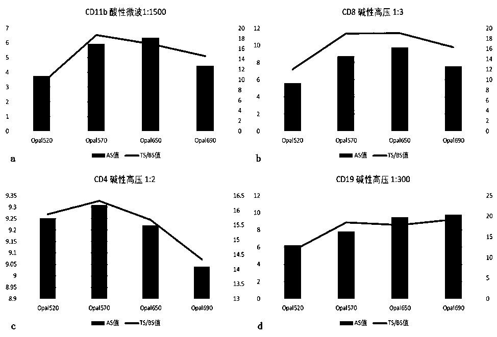
[0085] 1.1 Determine the optimal antigen retriev...

Embodiment 2

[0137] Example 2: The method of using the skin tissue in situ multi-marker immunostaining kit.

[0138] 2.1 Paraffin section pretreatment

[0139] Select paraffin sections of skin tissue and bake them at 60°C for 2 hours; take them out after 2 hours, soak them in preheated turpentine oil for 15 minutes, then soak them in 100% ethanol, 95% ethanol, 70% ethanol, and sterilized water for 10 minutes. minute.

[0140] 2.2 The first round of staining: CD11b-Opal570

[0141] 1) Take 200ml of acidic restoration solution (pH6.0), put it into an antigen restoration box containing slices, put the antigen restoration box into a microwave oven, adjust to high heat, and heat for 2-3 minutes (depending on the operating environment and usage The electric appliance brand is different, and the determination of the time depends on the observation of the boiling of the liquid surface in the repair box), when the repair liquid in the repair box starts to boil, quickly adjust to the low fire gear...

Embodiment 3

[0214] Example 3: Determination of detection indicators of skin tissue in situ multi-label immunostaining kit that can assist in the diagnosis and typing of lupus erythematosus

[0215] 3.1 Distinguish between normal people and lupus

[0216] 1) Select 18 cases of DLE, SLE, and SCLE skin lesions from different patients, and select 45 cases of normal human skin as a control, and use the skin tissue in situ multi-label immunostaining kit for sample detection to obtain corresponding data;

[0217] 2) Use IBM SPSS Statistics 23 to build a ROC analysis model, set the data of 54 lupus patients as the case group LE, and the data of 45 normal subjects as the normal control NC;

[0218] 3) CD4 single positive test results (CD4 / DAPI), CD8 single positive test results (CD8 / DAPI), CD19 single positive test results (CD19 / DAPI), CD11b single positive test results (CD11b / DAPI) , CD4-CD8 dual detection results ((CD4+CD8) / DAPI), CD4-CD19 dual detection results ((CD4+CD19) / DAPI), CD4-CD11b dua...


PUM


Abstract

The invention discloses a skin tissue immune cell in situ detection kit for typing lupus erythematosus and an application thereof. This kit uses in situ multi-label immunostaining and multi-spectral imaging technology to detect four molecular markers: CD4, CD8, CD19, and CD11b. The quadruple detection results of CD4‑CD8‑CD19‑CD11b can well distinguish discoid lupus erythematosus (DLE), subacute lupus erythematosus (SCLE), systemic lupus erythematosus (SLE) from healthy controls, combined with the single positive detection of CD19 Results and / or CD8-CD19 dual detection results, the accuracy rate is higher. The kit of the invention is simple in operation, economical and practical, and has high specificity for distinguishing different types of lupus erythematosus skin lesions, which facilitates its application and popularization in lupus erythematosus mechanism research and clinical diagnosis.

Description

technical field [0001] The invention belongs to the technical field of immune detection, in particular to a skin tissue immune cell in situ detection kit for typing lupus erythematosus and its application. Background technique [0002] Lupus erythematosus (Lupus erythematosus, LE) is a multifactorial chronic autoimmune disease that seriously affects the quality of life and endangers health. More than 80% of patients may develop skin involvement at the onset or progression. LE is a spectrum disease, one end is cutaneous lupus erythematosus (CLE), which presents a chronic benign course, and the lesions are mainly limited to the skin; the other end is systemic lupus erythematosus (SLE), which has multiple acute onset , the lesions can involve multiple systems, and severe cases can be life-threatening. Clinically, doctors divide LE into acute cutaneous lupus erythematosus (ACLE), subacute cutaneous lupus erythematosus (SCLE) and discoid erythematosus according to the appearanc...

Claims


Application Information

Patent Timeline
Patent Type & Authority Patents(China)
IPC IPC(8): G01N33/577G01N33/569
CPCG01N33/56966G01N33/577G01N2800/24
Inventor 陆前进吴海竞李倩文陈凯丽赵明胡凯
Owner THE SECOND XIANGYA HOSPITAL OF CENT SOUTH UNIV
Why Eureka
Try Eureka