Enteroscopy lymphoma auxiliary diagnosis system based on deep learning

An auxiliary diagnosis and deep learning technology, applied in image data processing, instrument, character and pattern recognition, etc., can solve problems such as high misdiagnosis rate, consumption of a lot of time, and delay in patient diagnosis time, so as to reduce workload and improve recognition. The effect of accuracy

Inactive Publication Date: 2020-05-01
SHANDONG UNIV QILU HOSPITAL +1
View PDF11 Cites 6 Cited by
  • Summary
  • Abstract
  • Description
  • Claims
  • Application Information

AI Technical Summary

Problems solved by technology

[0004] The small bowel endoscopy takes a long time, and the endoscopist is very prone to fatigue. At the same time, the endoscopist needs to spend a lot of time viewing the images during the withdrawal process of the small bowel scope. May cause missed or false detection of lesions due to physical fatigue and visual fatigue
[0005] In addition, due to the insidious onset of gastrointestinal lymphoma, atypical clinical symptoms and va

Method used


Image

  • Enteroscopy lymphoma auxiliary diagnosis system based on deep learning

Examples


Embodiment 1

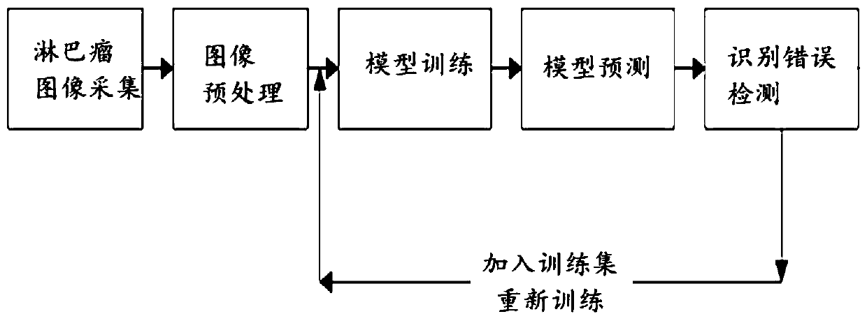
[0031] In one or more embodiments, a deep learning-based lymphoma auxiliary diagnosis system under enteroscopy is disclosed, including:

[0032] The image acquisition module is connected to the endoscope host through the acquisition card to obtain each frame of image information collected by the endoscope host;

[0033] The image preprocessing module is used to preprocess the collected image information; specifically includes:

[0034] Since the images are collected individually by the endoscope system in the clinic, it is necessary to remove the patient's private data in the images. In order to reduce the amount of calculation, it is necessary to remove the black frame and only keep the colored digestive tract area.

[0035] Through the black edge algorithm processing, zoom processing and normalization processing, each frame of the image is processed by the black edge algorithm to remove the redundant border of the endoscopic image and only retain the ROI area, and then use ...

Embodiment 2

[0049] In one or more embodiments, a terminal device is disclosed, which includes a processor and a computer-readable storage medium, the processor is used to implement instructions; the computer-readable storage medium is used to store multiple instructions, and the instructions are suitable for is loaded by the processor and executes the following processes:

[0050] Connect to the endoscope host through the acquisition card to obtain each frame of image information collected by the endoscope host;

[0051] Selecting a single frame of images with lymphoma lesions as a training sample, labeling the lesion area in the training sample, and generating labeling text information corresponding to the labeling position; the labeling area and the labeling text information corresponding to the area constitute a training set;

[0052] Construct an auxiliary diagnosis model, use the training set to optimize the training of the auxiliary diagnosis model; input the images to be detected i...


PUM


Abstract

The invention discloses an enteroscope lymphoma auxiliary diagnosis system based on deep learning. The system comprises an image collection module which is connected to an endoscope host through a collection card, and obtains the information of each frame of image collected by the endoscope host; the training set making module is used for selecting a single-frame image with lymphoma lesions as a training sample, labeling a lesion area in the training sample and generating labeling text information corresponding to a labeling position; the auxiliary diagnosis module is used for constructing anauxiliary diagnosis model and carrying out optimization training on the auxiliary diagnosis model by adopting the training set; and inputting an image to be detected into the trained auxiliary diagnosis model, and outputting an image classification result about whether lymphoma lesions exist or not. According to the method, the lymphoma lesion area under the enteroscope is automatically recognizedbased on the neural network algorithm, a doctor only needs to check the recognized image with lesion again, and the workload of picture check of the doctor is greatly reduced.

Description

technical field [0001] The invention belongs to the technical field of auxiliary intelligent diagnosis of lymphoma under enteroscopy, and in particular relates to an auxiliary diagnosis system of lymphoma under enteroscopy based on deep learning. Background technique [0002] The statements in this section merely provide background information related to the present invention and do not necessarily constitute prior art. [0003] Small intestinal lymphoma originates from lymphoid follicles under the small intestinal mucosa. Most intestinal lymphomas are a local manifestation of systemic lymphoma. Identifying the characteristics of the small intestinal mucosa through enteroscopy is the most effective method for the diagnosis of small intestinal lymphoma . [0004] The small bowel endoscopy takes a long time, and the endoscopist is very prone to fatigue. At the same time, the endoscopist needs to spend a lot of time viewing the images during the withdrawal process of the small...

Claims


Application Information

Patent Timeline
IPC IPC(8): G06T7/00G06K9/62
CPCG06T7/0012G06T2207/10068G06T2207/20081G06T2207/20084G06T2207/30028G06T2207/30096G06F18/241
Inventor 季锐杨笑笑冯建李延青辛伟邵学军左秀丽杨晓云李真
Owner SHANDONG UNIV QILU HOSPITAL
Why Eureka
Try Eureka