Blood vessel identification method based on SWI image and recurrent neural network
A technology of cyclic neural network and recognition method, applied in the field of nuclear magnetic resonance imaging, can solve problems such as inability to automatically distinguish, and achieve detailed and accurate results
- Summary
- Abstract
- Description
- Claims
- Application Information
AI Technical Summary
Problems solved by technology
Method used
Image


Examples
Embodiment 1
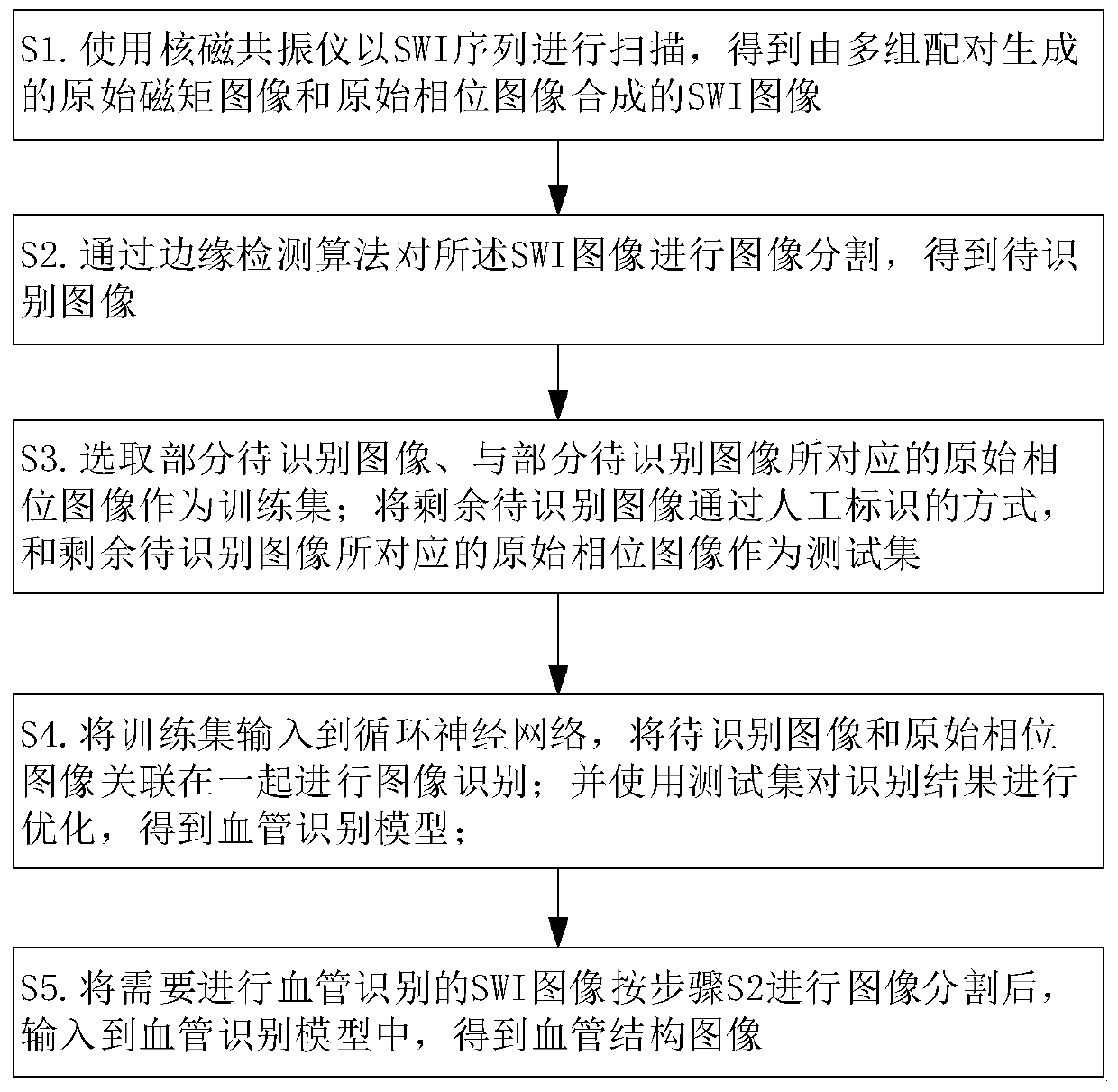
[0029] The invention provides a blood vessel recognition method based on SWI image and cyclic neural network, comprising the following steps:
[0030] S1. Use the nuclear magnetic resonance instrument to scan with the SWI sequence to obtain the original magnetic moment image and the original phase image generated by multiple pairs. The original magnetic moment image and the original phase image are fused after image post-processing to obtain the SWI image;
[0031] S2. Segmenting the SWI image through an edge detection algorithm to obtain an image to be recognized;
[0032] S3. Selecting part of the images to be recognized and the original phase images corresponding to the part of the images to be recognized as a training set; using the remaining images to be recognized by manual marking, and the original phase images corresponding to the remaining images to be recognized as a test set;
[0033] S4. Input the training set into the cyclic neural network, associate the image to ...
Embodiment 2
[0064] Judging from the physiological and anatomical images of the human body, blood vessels have their structural characteristics. The overall structure of blood vessels is tree-like with multiple branches; the local structure is tubular with a certain curvature. In the SWI image, if the tissue shows low signal, and at the same time it is tree-like as a whole and has multiple branches, it means that the tissue is a venous vessel.
[0065] On the technical scheme of embodiment 1, change. The SWI image is obtained by scanning with a nuclear magnetic resonance apparatus, and the image segmentation is performed on the SWI image to obtain the image to be recognized. These two steps refer to the technical solution of Embodiment 1. From the point of view of human anatomy section, the SWI image scanned by MRI is a group of layered, continuous, multiple two-dimensional images; on the space coordinate axis of the section, the distance between layers is 0.5-1mm . When carrying out th...
Embodiment 3
[0068] An MRI machine is used to scan with a SWI sequence, and usually images of veins are shown on the images. However, in the actual work of medical personnel, it is often necessary to observe both venous blood vessels and arterial blood vessels at the same time.
[0069] In order to solve the above-mentioned technical problems, on the basis of Embodiment 1 and Embodiment 2, the technical solution adopted is: when using the nuclear magnetic resonance instrument to scan with the SWI sequence, apply an inclination angle to the second echo of the SWI, Three-dimensional fully flow-compensated arterial angiography was performed. In this way, arterial vessels and venous vessels can be clearly imaged at the same time, and a SWI image that clearly displays both arterial vessels and venous vessels on the same image can be obtained. Preferably, the applied inclination angle is 15 degrees to 25 degrees.
[0070] In this way, for the SWI image that clearly shows arterial blood vessels...
PUM
 Abstract
Description
Claims
Application Information
 - R&D
- Intellectual Property
- Life Sciences
- Materials
- Tech Scout
- Unparalleled Data Quality
- Higher Quality Content
- 60% Fewer Hallucinations
Browse by: Latest US Patents, China's latest patents, Technical Efficacy Thesaurus, Application Domain, Technology Topic, Popular Technical Reports.
© 2025 PatSnap. All rights reserved.Legal|Privacy policy|Modern Slavery Act Transparency Statement|Sitemap|About US| Contact US: help@patsnap.com