Anti-EGFR/PD-1 bispecific antibody

A bispecific antibody, PD-1 technology, applied in the direction of antibodies, specific peptides, anti-tumor drugs, etc.

Active Publication Date: 2020-05-26
SUNSHINE GUOJIAN PHARMA (SHANGHAI) CO LTD
View PDF8 Cites 3 Cited by
  • Summary
  • Abstract
  • Description
  • Claims
  • Application Information

AI Technical Summary

Problems solved by technology

Antibody combinations often do not have this effect

Method used


Image

  • Anti-EGFR/PD-1 bispecific antibody
  • Anti-EGFR/PD-1 bispecific antibody
  • Anti-EGFR/PD-1 bispecific antibody

Examples


Embodiment 1

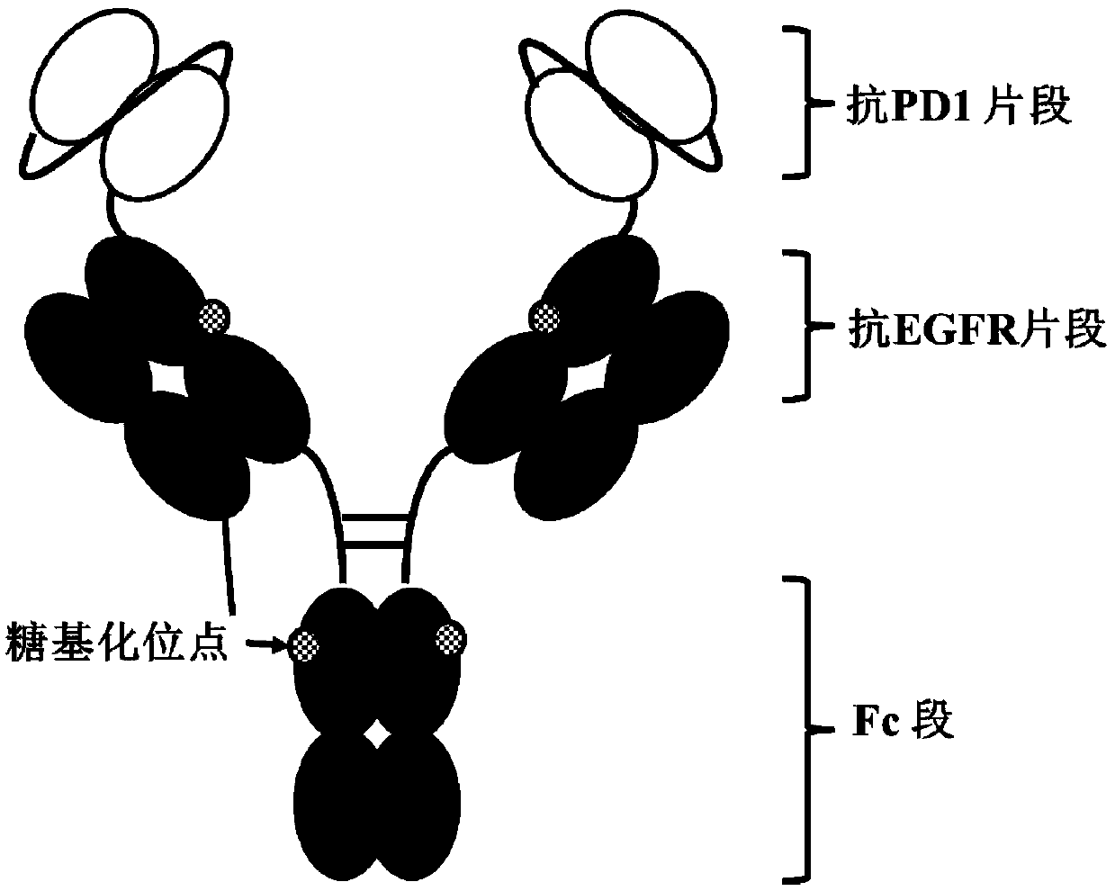
[0122] Example 1 Molecular construction of anti-EGFR / PD-1 bispecific antibody

[0123] In the present invention, the anti-EGFR / PD-1 bispecific antibody is constructed by using scFv and IgG in series, and the specific form is scFv-L2-IgG, such as figure 1 shown. Among them, the scFv adopts the molecular form of VH-L1-VL. The heavy chain variable region VH (SEQ ID NO: 13) and the light chain variable region VL (SEQ ID NO: 14) of the anti-PD-1 monoclonal antibody are connected through L1 (SEQ ID NO: 17) to obtain the anti-PD-1 monoclonal antibody Single-chain antibody fragment scFv of PD-1 (SEQ ID NO: 19). Using L2 (SEQ ID NO: 18) to link the single-chain antibody fragment with the heavy chain of the anti-EGFR monoclonal antibody to obtain the heavy chain of the anti-EGFR / PD-1 bispecific antibody molecule (SEQ ID NO: 20) , the light chain of the EGFR monoclonal antibody (SEQ ID NO: 21) remained unchanged. In order to improve the expression efficiency of antibody molecules in ...

Embodiment 2

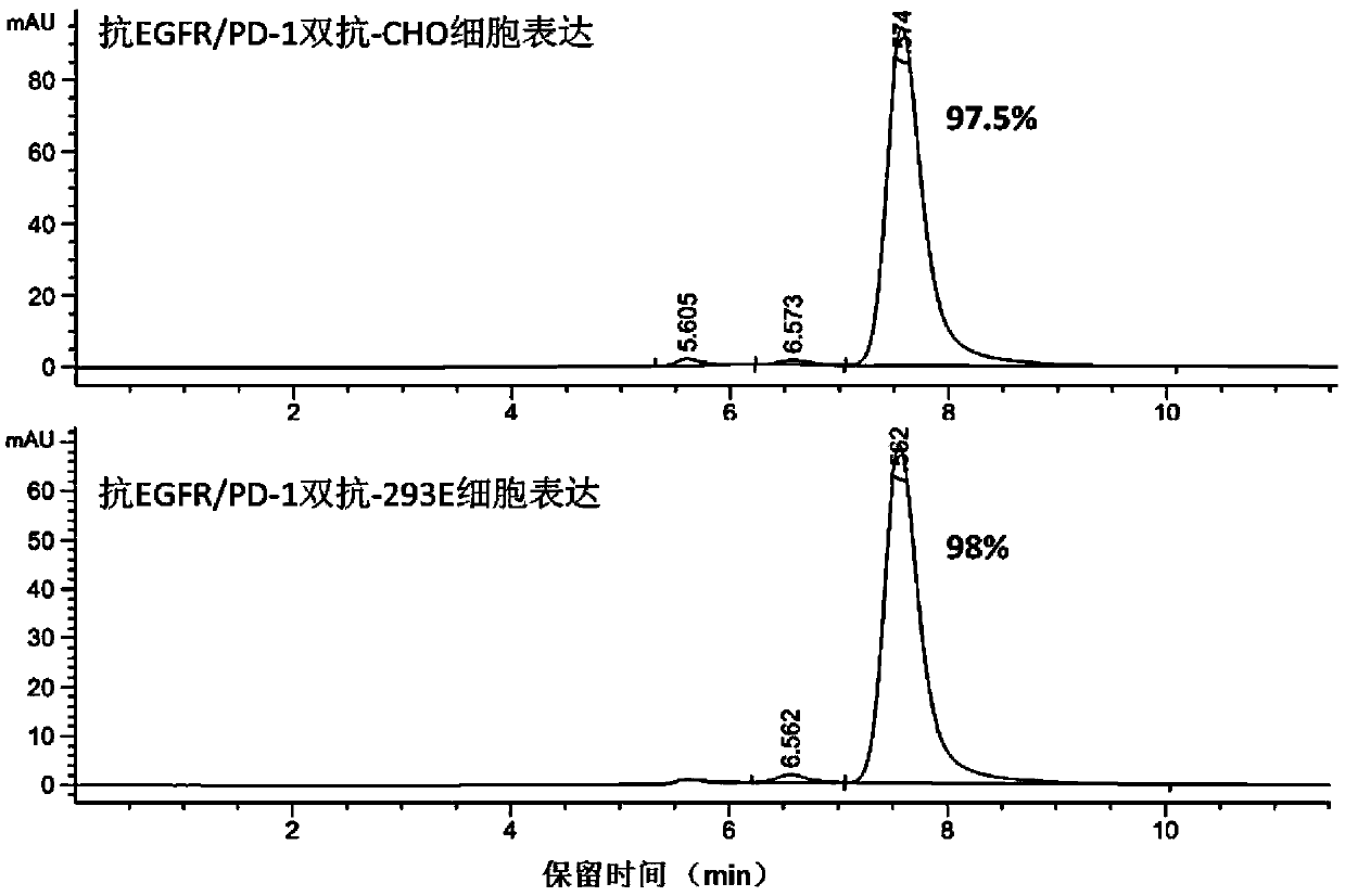
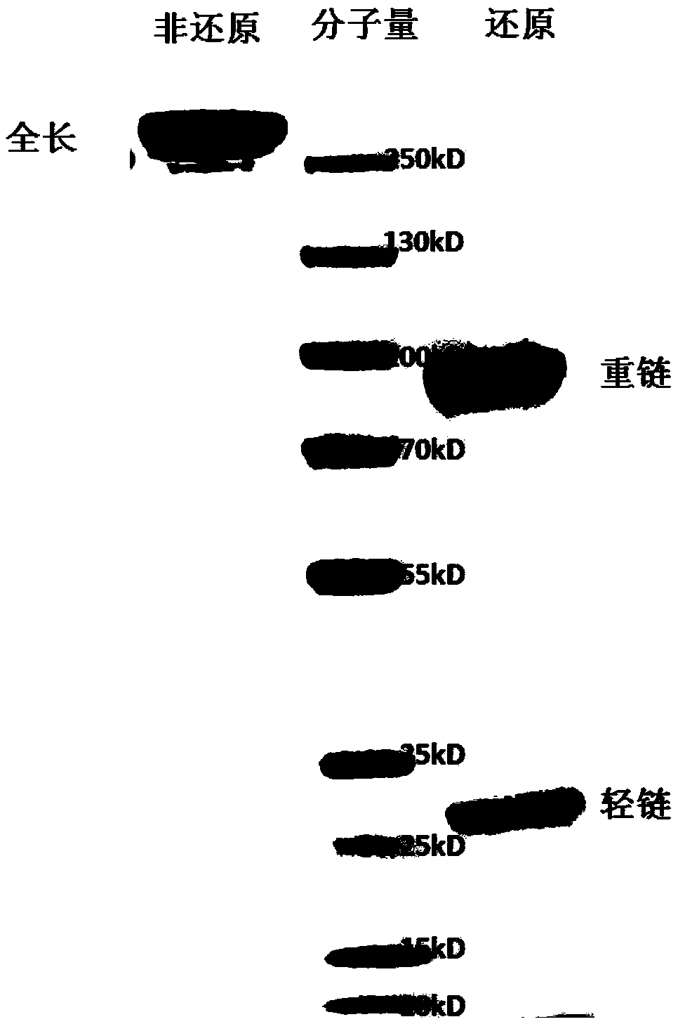
[0124] Example 2 Expression and purification of anti-EGFR / PD-1 bispecific antibody

[0125] The DNA fragments of the heavy chain and light chain of the anti-EGFR / PD-1 bispecific antibody were subcloned into the pTT5 vector, and the recombinant plasmids were extracted and co-transfected into CHO cells and / or 293E cells. After 7 days of cell culture, the culture solution was subjected to high-speed centrifugation and vacuum filtration with a microporous membrane, and then loaded onto a HiTrap MabSelectSuRe column, and the protein was eluted in one step with 100mM citric acid, pH3.5 eluent, and the target sample was recovered and dialyzed. solution to PBS. The purified protein was detected by HPLC, such as Figure 2A As shown, the molecular state of the antibody is uniform, and the purity of the monomer reaches more than 97%. Add the reduced protein electrophoresis loading buffer and the non-reduced protein electrophoresis loading buffer respectively, and detect after boiling. ...

Embodiment 3

[0126] Example 3 Determination of affinity of anti-EGFR / PD-1 bispecific antibody to antigen by enzyme-linked immunosorbent assay (ELISA)

[0127] 3.1 ELISA to detect the affinity of anti-EGFR / PD-1 bispecific antibody to EGFR

[0128] Dilute the recombinant EGFR-ECD-Fc protein with coating solution to 3 μg / ml, add 50 μl / well to the microtiter plate, and overnight at 4°C. Wash the plate 3 times with PBST, add 200 μl / well blocking solution, place at 37°C for 1 hour, wash the plate once with PBST and set aside. Dilute the anti-EGFR / PD-1 bispecific antibody to 100 μg / ml with diluent, form 12 concentration gradients (the highest concentration is 100,000 ng / ml, the lowest concentration is 0.02 ng / ml), and add the blocked ELISA plate, 100 μl / well, place at 37°C for 1 hour. Wash the plate three times with PBST, add HRP-labeled mouse anti-human Fab antibody, and place at 37°C for 30 minutes. After washing the plate with PBST for 3 times, pat dry the remaining droplets on absorbent pa...


PUM


Abstract

The invention belongs to the field of tumor treatment, and discloses an anti-EGFR / PD-1 bispecific antibody, a preparation method and an application in tumor resistance. More specifically, a single-chain variable fragment scFv and an immunoglobulin antibody IgG are connected through a peptide joint L2 to obtain the bispecific antibody with a structure and a function similar to those of a full antibody; the bispecific antibody has the functions of targeting a tumor surface antigen EGFR and activating tumor immunity, and can effectively exert the ADCC function of an Fc segment of the antibody. Compared with a monoclonal antibody, the molecule has an obvious synergistic effect, and the anti-tumor effect of the molecule is superior to that of antibody combination.

Description

technical field [0001] The invention belongs to the field of tumor treatment and biotechnology, and relates to a preparation method and application of a bispecific antibody molecule against EGFR and PD-1. Background technique [0002] EGFR (Epidermal Growth Factor Receptor) is a receptor for epidermal growth factor (EGF), which belongs to the ErbB receptor family. EGFR is a transmembrane glycoprotein with a molecular weight of 170KDa, which belongs to receptor-type tyrosine kinase. Under the action of related ligands such as EGF and transforming growth factor-α (transforming growth factorα, TGFα), EGFR is converted from monomer Activated for dimerization, thereby further activating downstream signaling pathways and regulating cell proliferation. A large number of studies have shown that there are high or abnormal expressions of EGFR in most tumors such as glial cell carcinoma, renal carcinoma, lung cancer, prostate cancer, pancreatic cancer, breast cancer and other tissues....

Claims


Application Information

Patent Timeline
IPC IPC(8): C07K16/46C12N15/13A61K39/395A61P35/00
CPCC07K16/2818C07K16/2863A61P35/00C07K2317/31C07K2317/622C07K2317/565C07K2317/92C07K2317/76C07K2317/732A61K2039/505C07K16/46A61K39/395
Inventor 朱祯平黄浩旻李理
Owner SUNSHINE GUOJIAN PHARMA (SHANGHAI) CO LTD
Why Eureka
Try Eureka