Antibody resisting lymphocyte activation gene 3 (LAG-3) and application
A technology of lymphocytes and genes, applied in the direction of antibodies, applications, anti-receptors/cell surface antigens/cell surface determinant immunoglobulins, etc., can solve the problems of lack of antibodies and achieve strong binding activity and excellent synergistic effects
- Summary
- Abstract
- Description
- Claims
- Application Information
AI Technical Summary
Problems solved by technology
Method used
Image

Examples
Embodiment 1
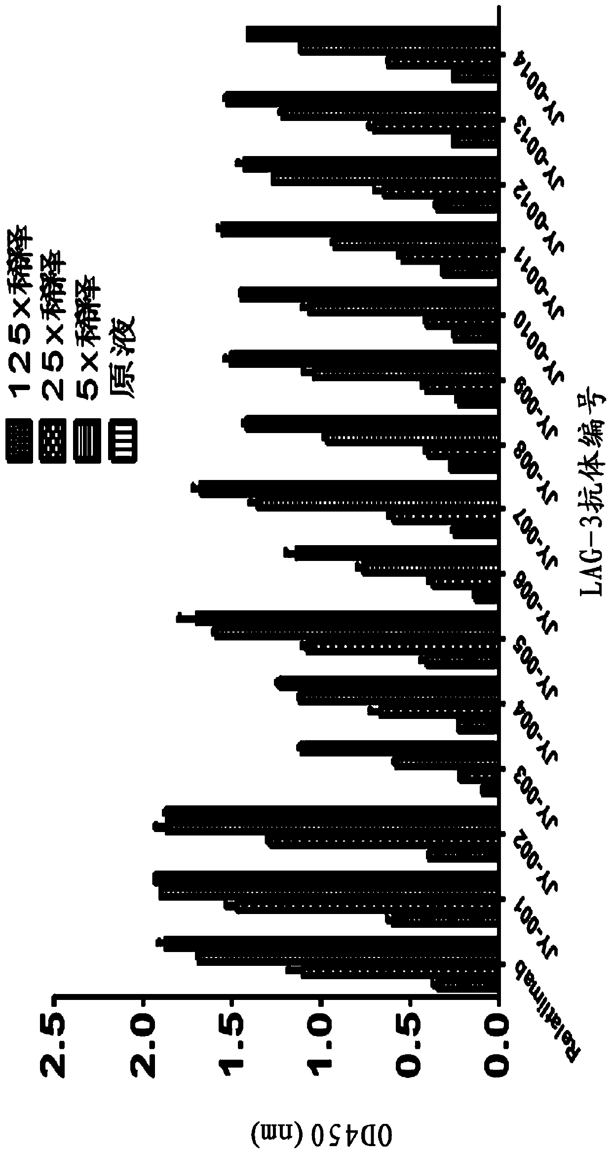
[0034] Example 1: Screening of Anti-LAG-3 Antibody from Natural Phage Antibody Library
[0035] 1.1 Materials
[0036] Reagents: ampicillin, kanamycin, yeast extract, glucose, Tween 20, TMB, PBS, PEG, etc. were routine reagents, and anti-M13 HRP secondary antibody was purchased from Amersham Company.
[0037] Bacterial strains and phagemids: TG1 Escherichia coli strain and helper phage VCSM13 are commercial products.
[0038] 1.2 Method
[0039] 1.2.1 Antigen-specific phage display antibody screening
[0040] 1) Dilute the corresponding LAG-3 / Fc antigen to 10 μg / mL in sterile PBS, add 500 μL per tube into the immunotube, and coat overnight at 4°C.
[0041] 2) Discard the antigen solution, add 4% milk prepared in sterile PBST, and block for one hour at room temperature.
[0042] 3) Discard the milk, add 500 μL of blocked phage solution to the immunotube, and incubate at room temperature for one hour
[0043] 4) Wash the tube 15 times with PBST, and then wash the tube 10 ti...
Embodiment 2
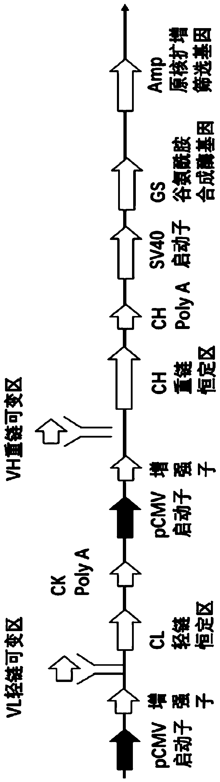
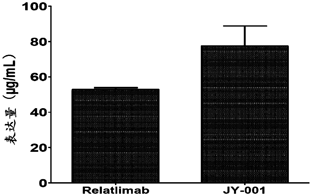
[0071] Example 2: Construction and expression of anti-LAG-3 full-length antibody JY-001 expression vector
[0072] 2.1 Materials
[0073] Primers were synthesized by Sangon Bioengineering (Shanghai) Co., Ltd.; Q5 DNA polymerase was a product of NEB Company; T4 DNA ligase was a product of NEB Company; gene synthesis and Jetprime transfection reagent were purchased from French Polyplus Company; sequencing was provided by Sangon Bioengineering ( Shanghai) Co., Ltd.; other related reagents are commercially available.
[0074] 2.2 Method
[0075] 2.2.1 Construction of anti-LAG-3 full-length antibody expression vector
[0076] The light and heavy chain variable region sequence of the JY-001 antibody was obtained through Example 1, and the JY-001 variable region gene was cloned into the eukaryotic expression vector pJY-GS / KIgG4 containing the human IgG4 heavy chain constant region and the kappa light chain constant region , to construct the whole antibody expression vector, its ph...
Embodiment 3
[0084] Example 3: Analysis of Anti-LAG-3 Antibody Binding Activity
[0085] 3.1 Materials
[0086] LAG-3 / Fc antigen was purchased from Beijing Yiqiao Shenzhou Technology Co., Ltd.; goat anti-human Fab-HRP (horseradish peroxidase-labeled secondary antibody) was purchased from abcam company in the United States; casein, PBS, H 2 SO 4 For routine experimental reagents.
[0087] 3.2 Method
[0088] Dilute the LAG-3 / Fc antigen to 1 μg / mL in the coating solution, add 100 μL per well to the enzyme-linked plate, and place it in a humid box at 4°C overnight. Wash the enzyme-linked plate 3 times with a plate washer, block with 1.5% casein, 200 μL per well, and block for 1 hour at 37°C in a humid box. Dilute the LAG-3 antibody to 10 μg / mL with 1×PBS, and dilute it in a 2-fold gradient, add 100 μL per well to the enzyme-linked plate, react in a wet box at 37°C for 1 hour, wash the enzyme-linked plate 3 times, add sheep Anti-human Fab-HRP secondary antibody reacted at room temperature...
PUM
 Abstract
Description
Claims
Application Information