Localization and fixation device for animal tibia surgery

A surgical operation and fixation device technology, applied in veterinary surgery, animal restraint equipment, medical science, etc., can solve the problems of tibial curvature, small size, and limited surgical operation space

Active Publication Date: 2020-06-05
王力平
View PDF6 Cites 3 Cited by
  • Summary
  • Abstract
  • Description
  • Claims
  • Application Information

AI Technical Summary

Problems solved by technology

Despite unremitting efforts, a novel ultra-thin cannulated bone fixation needle was invented, which realized the possibility of electrically driving percutaneous bone fixation needles for small animal bones. However, the shape of the mouse tibia is curved and the size is extremely large Small size, limited surgical operation space, and the anesthetized mouse body is soft, and the anatomical part of the bone under the fur and soft tissue is d

Method used


Image

  • Localization and fixation device for animal tibia surgery
  • Localization and fixation device for animal tibia surgery
  • Localization and fixation device for animal tibia surgery

Examples


Embodiment 1

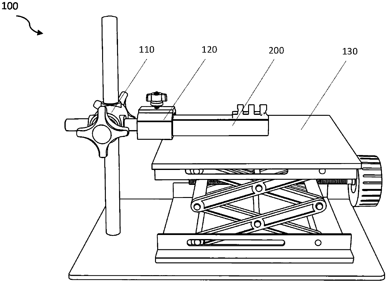
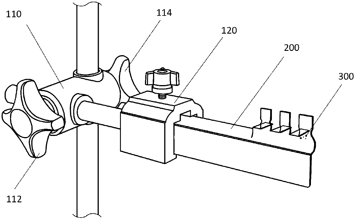
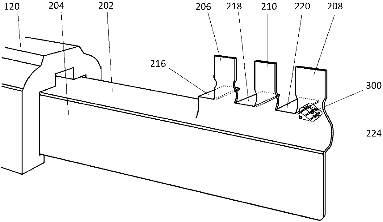
[0070] like Figure 1-10 The animal tibia positioning and fixing device 100 shown includes a tibial positioning and fixing arm 200 connected to an external fixation frame, and the lifting operating table 130 is used in conjunction with the animal tibia positioning and fixing device 100 . The external fixing frame is a three-dimensional adjustable fixing frame. The three-dimensional three-dimensional adjustable fixing frame is composed of a horizontal axis, a vertical axis and a three-dimensional three-dimensional controller 110 connecting the horizontal axis and the vertical axis. The tibial positioning fixation arm 200 is connected with the horizontal axis connected to the three-dimensional three-dimensional adjustable fixing frame through the surgical positioning trimmer 120 . The tibial positioning and fixing arm 200 is provided with a popliteal fossa positioning unit, a knee fixing unit and an ankle fixing unit. The tibial surgical positioning and fixing arm is used to de...


PUM


Abstract

The invention discloses a localization and fixation device for animal tibia surgery. The localization and fixation device comprises a tibia localization and fixation arm connected with an external fixation frame; the tibia localization and fixation arm is provided with a popliteal fossa localization unit, a knee fixation unit and an ankle fixation unit; and the tibia localization and fixation armis used for defining and fixing a surgery standard position of an animal tibia during tibia surgery, and thus, a surgical small environment operating platform of a three-dimensional standard positionimage of the tibia surgery is formed. According to the localization and fixation device, three-dimensional fixed-point surgical fixation for the orthopedic surgery on a laboratory animal tibia is realized, and thereby, implementation of tibia bone fixation needle puncture, the surgical standardized operation of a fracture and segmental bone defect experimental model, and the repeatability and thedata reliability of a surgical technique of an experimental animal tibia model can be guaranteed. Assisted by a three-dimensional adjustable fixation frame as the external fixation frame, the localization and fixation device makes the tibia localization and fixation arm have stable three-dimensional variability based on vertical and horizontal axial directions, so that a required surgical big environment suitable body position can be provided for a surgery operator.

Description

technical field [0001] The invention relates to a special surgical instrument for medical experimental animal models, specifically for the establishment of experimental animal models of tibial fractures and tibial segmental bone defects, and the implementation of orthopedic external fixation surgery techniques. Orthopedic surgery positioning fixation device for repeatability, bone fixation needle percutaneous puncture anchoring for tibial external fixation, fixed-point digital surgery for tibial fractures or segmental bone defects. Background technique [0002] At present, the external fixation technology of the small animal tibia experimental model in the international laboratory has always been a technical problem that many laboratories can't expect. Due to the difficulty in implementing standard and repeatable small animal bone surgery with conventional surgical instruments and general laboratory technical background, many laboratories around the world have not been able ...

Claims


Application Information

Patent Timeline
IPC IPC(8): A61D1/00A61D3/00
CPCA61D1/00A61D3/00A61D2003/006
Inventor 王力平
Owner 王力平
Why Eureka
Try Eureka