Animal femoral surgery positioning and fixing frame

A fixed frame, femur technology, applied in the direction of animal restraint equipment, medical science, veterinary surgery, etc., to achieve the effect of improving operability

Active Publication Date: 2021-04-02
HANGZHOU HUAMAI MEDICAL DEVICES CO LTD
View PDF11 Cites 0 Cited by
  • Summary
  • Abstract
  • Description
  • Claims
  • Application Information

AI Technical Summary

Problems solved by technology

[0010] However, this device is only suitable for animal tibial surgery, so it is necessary to develop a three-dimensional animal femur positioning and fixing device, focusing on the operability of small animal femur surgery, standardization, repeatability, and standardization of laboratory animal surgery requirements. Reliability is implemented in the technical procedures of controlled operation, and finally reflected in the operation results of animal experimental models

Method used


Image

  • Animal femoral surgery positioning and fixing frame
  • Animal femoral surgery positioning and fixing frame
  • Animal femoral surgery positioning and fixing frame

Examples


Embodiment 1

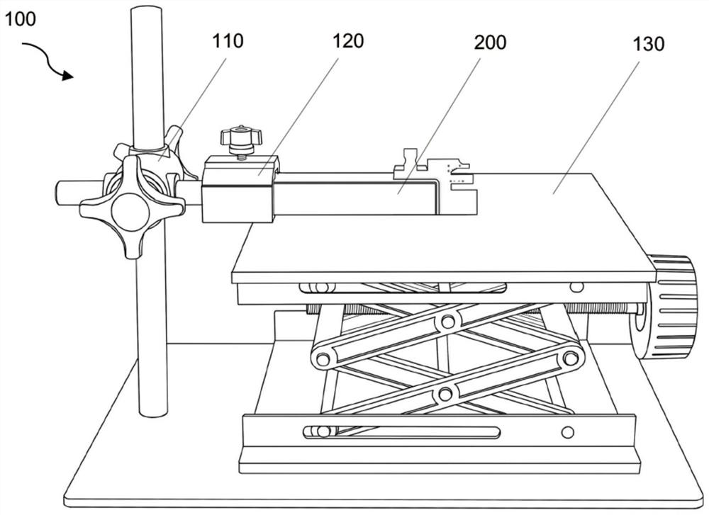
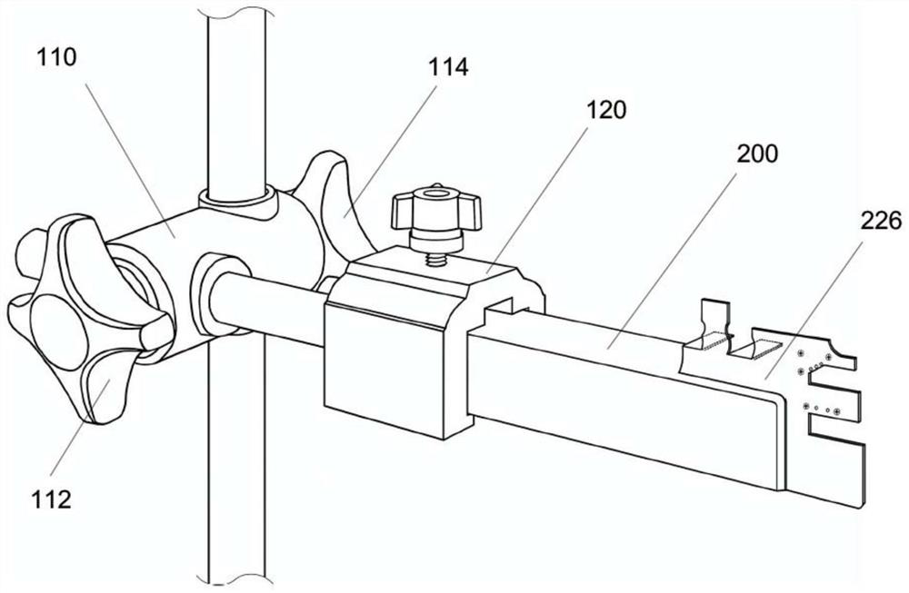
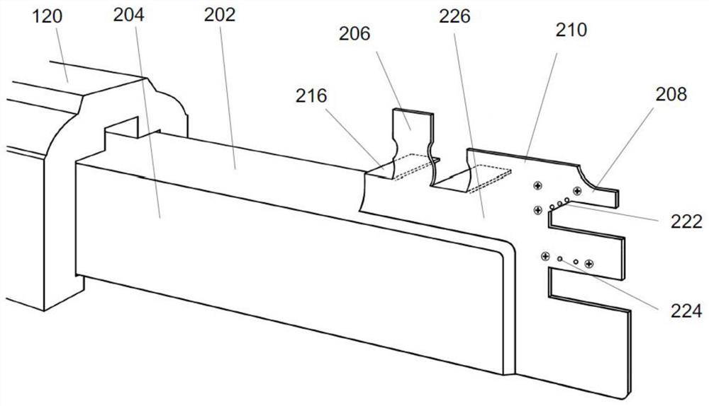
[0087] like Figure 1-2 The animal femur surgery positioning fixture 100 shown includes a femoral positioning fixation arm 200 connected to an external fixator, and the lifting operating table 130 is used in conjunction with the animal femur surgery positioning fixture 100 . The external fixing frame is a three-dimensional adjustable fixing frame. The three-dimensional three-dimensional adjustable fixing frame is composed of a horizontal axis, a vertical axis and a three-dimensional three-dimensional controller 110 connecting the horizontal axis and the vertical axis. Femoral shaft positioning and fixing unit, popliteal fossa positioning unit, knee fixing unit and ankle fixing unit are provided on the femoral positioning and fixing arm 200. The femoral surgery positioning and fixing arm is used to limit and fix the standard position of the animal femur in femoral surgery. A small operating environment operation platform for three-dimensional standard image of femur surgery is...

Embodiment 2

[0125] like Figure 32 Shown, is the use of rigid external fixator (the so-called rigid external fixator is relative to Figure 33 -Schematic diagram of mouse femur fracture surgery for the flexible external fixator of Example 3). Six needles penetrate the mouse femur and anchor the root in place; three needles at each end of the femur have outer segments bent parallel to each other toward the center to form shoulder bridges; fractures of the femur are generated with a bone breaker; coated with light-curable flowable composite The bridge is filled; the composite is cured with LED lights; the cured portion of the external fixator is removed after a few weeks by cutting the pins; all remaining pins are then spun out.

Embodiment 3

[0127] like Figure 33 Shown is a schematic diagram of mouse femur fracture surgery using a flexible external fixator. Six needles penetrated the mouse femur and anchored in place at the root; three distal femoral needles and three proximal femoral needles were each bent toward each other with three needles at each end in parallel to form two end cluster bridges; Each of them is coated with a light-curable flowable composite material and cured with an LED light; two elastic pins are placed and the two clusters at the two ends are connected step by step, that is, each left end and each right end of the two ends are connected and cured step by step through a light-curable flowable composite material. point; the right elastic pin is used to connect the right end faces of the two clusters, but only the proximal end is cured temporarily; the left elastic pin is used to connect the left end faces of the two clusters, but only the distal end is cured temporarily; the two elastic pins...


PUM


Abstract

The invention discloses an animal femoral surgery positioning and fixing frame which comprises a femoral positioning and fixing arm. The femoral positioning and fixing arm is connected to an externalfixing frame and provided with a popliteal space positioning unit, a femoral shaft positioning and fixing unit, an ankle fixing unit and a knee fixing unit; and a femoral shaft positioning needle, a femoral shaft positioning needle clip shaped like a Chinese character 'hui' and a femoral shaft positioning needle base in the femoral shaft positioning and fixing unit jointly form an elastic clampingring for positioning and fixing a femoral shaft. According to the animal femoral surgery positioning and fixing frame, the animal femur is positioned and fixed to the femoral positioning and fixing arm through four sites (the femoral shaft, the popliteal space, the knee and the ankle) in anatomy, the femoral surgery positioning and fixing arm limits and fixes the surgery standard position of a femoral entity object in a surgery, a surgery microenvironment operation platform of a femoral surgery three-dimensional standard position image is formed, and therefore surgery standardized operation of femoral bone fixing needle puncture, fracture and segmental bone defect experiment models, repeatability of the experiment animal femoral surgery and data reliability are guaranteed.

Description

technical field [0001] The invention relates to a special surgical instrument for a medical experimental animal model, specifically for the establishment of experimental animal models of femoral fractures and femoral segmental bone defects, and the implementation of femoral external fixation surgery techniques. Orthopedic surgery positioning and fixing device for repeatability, percutaneous puncture anchoring of bone fixation pins to ensure femoral external fixation, fixed-point digital surgery for femoral fractures or segmental bone defects. Background technique [0002] Complications associated with the treatment of long bone fractures remain a major challenge for orthopedic surgeons. Elucidating the mechanisms of bone healing and development, and subsequently altering these mechanisms to improve outcomes, often requires animal models as an intermediary between in vitro and human clinical studies. The mouse femur model is the most commonly used model in translational rese...

Claims


Application Information

Patent Timeline
Patent Type & Authority Applications(China)
IPC IPC(8): A61D3/00A61D1/00
CPCA61D3/00A61D1/00A61D2003/006
Inventor 王力平江涛
Owner HANGZHOU HUAMAI MEDICAL DEVICES CO LTD
Why Eureka
Try Eureka