
Device for interventional therapy of adult congenital heart disease under guidance of echocardiogram

A congenital heart disease, echocardiography technology, applied in the direction of guide wire, medical science, surgery, etc., to achieve the effect of reducing the perforation rate and improving safety

Pending Publication Date: 2020-06-12
JIANGSU PROVINCE HOSPITAL THE FIRST AFFILIATED HOSPITAL WITH NANJING MEDICAL UNIV
View PDF0 Cites 1 Cited by
  • Summary
  • Abstract
  • Description
  • Claims
  • Application Information

AI Technical Summary

Problems solved by technology

Due to the above problems, only a few top three hospitals have been able to routinely apply this technology, and most hospitals have not been able to routinely carry out this technology

Method used


Image

  • Device for interventional therapy of adult congenital heart disease under guidance of echocardiogram
  • Device for interventional therapy of adult congenital heart disease under guidance of echocardiogram
  • Device for interventional therapy of adult congenital heart disease under guidance of echocardiogram

Examples


Embodiment 1

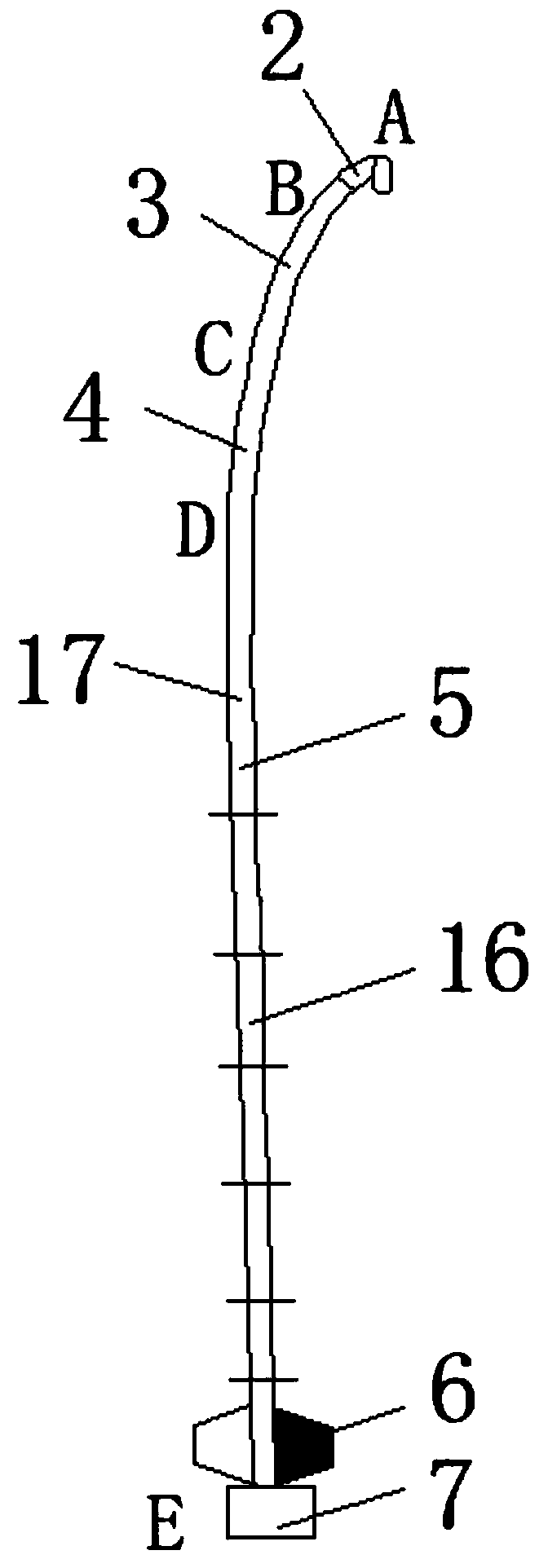
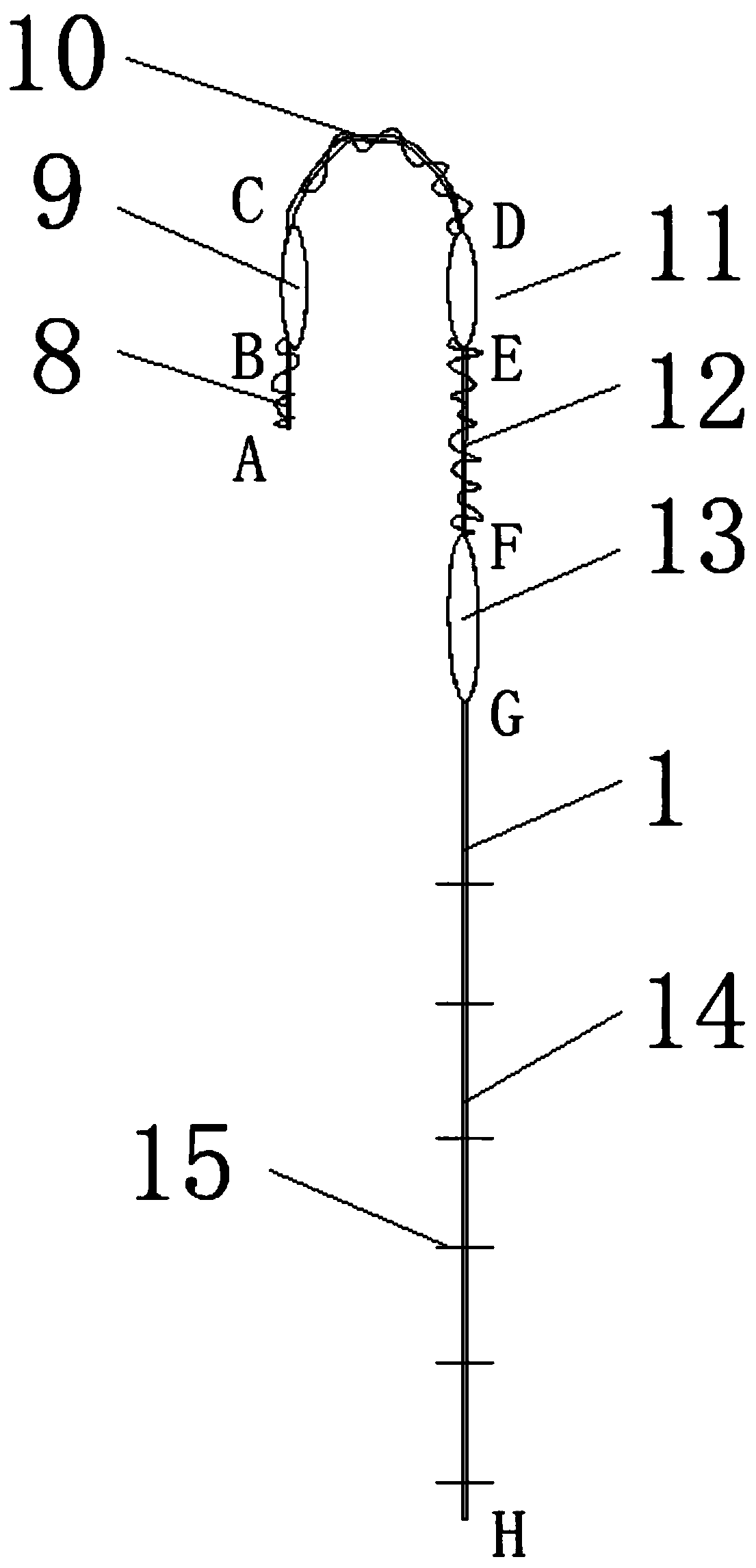
[0034]Embodiment 1: Use the right heart scale catheter 5 to measure the distance from the third intercostal space on the right midclavicular line to the venipuncture point, as the working distance, place the femoral vein sheath in the punctured femoral vein, and the head of the routing guide wire 1 should extend out of the right The outer 3cm of the heart scale catheter 5, push the right heart scale catheter 5 and the routing guide wire 1 forward together, for those who pass through the femoral vein, the inferior vena cava can be displayed on the cut plane below the xiphoid process, and the right heart scale catheter 5 and the routing can be monitored Guidewire 1 passing condition. After the right heart graduated catheter 5 and routing guide wire 1 are inserted into the body to reach the working distance, the routing guide wire 1 is withdrawn, the right heart graduated catheter 5 is gently rotated, and the right heart graduated catheter 5 can be found on the four-chamber view b...

example 2

[0035] Example 2: Use the right heart graduated catheter 5 to measure the distance from the third intercostal space at the right border of the sternum to the puncture point of the right femoral vein, and use it as the working distance for the right heart graduated catheter 5 to operate. Insert the vascular sheath through the femoral vein, and send the routing guide wire 1 and the right heart graduated catheter 5 through the vascular sheath. When the right heart graduated catheter 5 enters the body and reaches the working distance, the routing guide wire 1 is appropriately withdrawn, and the direction of the head end of the right heart graduated catheter 5 is gently rotated to facilitate ultrasonic exploration of the position of the catheter in the right atrium. Guide wire and catheter from the right atrium through the tricuspid valve into the right ventricle. Slightly retreat the routing guide wire 1, use the operation wing 6 to adjust the catheter direction, make the opening ...


PUM

PropertyMeasurementUnit
Lengthaaaaaaaaaa
Lengthaaaaaaaaaa
Lengthaaaaaaaaaa

Abstract

The invention discloses a device for interventional therapy of adult congenital heart disease under the guidance of echocardiogram. The device comprises a path finding guide wire and a right heart scale catheter, and the path finding guide wire comprises a guide wire AB section, a guide wire BC section, a guide wire CD section, a guide wire DE section, a guide wire EF section, a guide wire FG section and a guide wire GH section. By arranging the path finding guide wire and guide wire scales, the probability of entering small vessels along the path can be remarkably reduced by adopting a U-shaped guide wire head. When the path finding guide wire is withdrawn to the middle of the right heart scale catheter, the right heart scale catheter can change the advancing direction of the path findingguide wire through rotation, and therefore it is guaranteed that the path finding guide wire safely passes through inferior vena cava, superior vena cava, atrial septum, atrioventricular valve, pulmonary artery and other parts and reaches a preset position under echocardiogram monitoring. Conditions are created for follow-up of the matched right heart scale catheter to a target site and measurement of the length of the matched right heart scale catheter entering the body, and it is in a safe path that the matched right heart scale catheter is pushed out of a purple area of the routing guide wire along the routing guide wire.

Description

technical field [0001] The invention relates to the technical field of heart disease treatment devices, in particular to a device for interventional treatment of adult congenital heart disease guided by echocardiography. Background technique [0002] Traditional percutaneous interventional techniques are generally completed in the catheterization room under the guidance of radiation, and most patients need to use contrast media, which will bring potential radiation damage to patients and operators, and the use of contrast media has the risk of renal function damage and allergy; Moreover, the operator needs to wear heavy protective equipment, which not only brings inconvenience to the operation, but also increases the labor intensity of the operator. [0003] In 2000, Ewert et al. reported for the first time that ASD closure was performed under the guidance of transesophageal echocardiography (TEE) without X-ray fluoroscopy. Chinese Expert Consensus. Due to the separation f...

Claims


Application Information

Patent Timeline
IPC IPC(8): A61B17/00A61M25/09
CPCA61B17/0057A61B17/00234A61M25/09A61B2017/00243A61M2025/09066A61B2017/00623
Inventor 王亮孔祥清袁振茂杨荣盛燕辉孙伟张浩
Owner JIANGSU PROVINCE HOSPITAL THE FIRST AFFILIATED HOSPITAL WITH NANJING MEDICAL UNIV
Learn More