Automatic splicing method and system for ABUS mammary gland ultrasonic panorama and storage medium

An automatic stitching and panorama technology, which is applied to the details of image stitching, image enhancement, image analysis, etc., can solve the problems of limited field of view and inability to fully present the mammary gland area, and achieve high automation, high flatness, and smooth operation process. simple effect

Active Publication Date: 2020-06-12
YUNNAN UNIV
View PDF19 Cites 2 Cited by
  • Summary
  • Abstract
  • Description
  • Claims
  • Application Information

AI Technical Summary

Problems solved by technology

Automatically splicing flat and uniform ABUS breast ultrasound panoramic images to solve the problem that the

Method used


Image

  • Automatic splicing method and system for ABUS mammary gland ultrasonic panorama and storage medium
  • Automatic splicing method and system for ABUS mammary gland ultrasonic panorama and storage medium
  • Automatic splicing method and system for ABUS mammary gland ultrasonic panorama and storage medium

Examples


Embodiment 1

[0056] This embodiment discloses an automatic splicing method for ABUS breast ultrasound panoramas, which can splice coronal ultrasound images (C-plane ultrasound images) at different scanning positions and at the same scanning depth into a panoramic image. Splicing methods include:

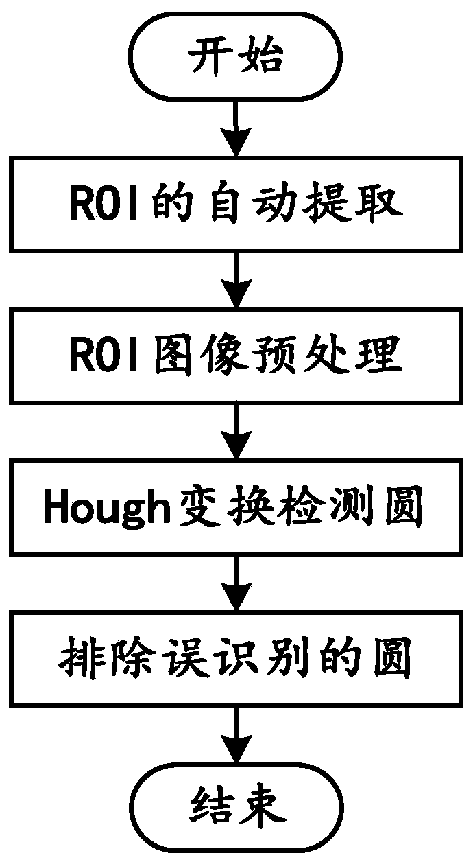
[0057] For each ultrasonic image in the ultrasonic image set to be spliced, the position of the feature point is respectively located, and the position of the feature point is used as a matching feature point for splicing and exists in each ultrasonic image. Generally speaking, in order to facilitate accurate identification, nipples are selected as feature points. Taking the nipple as a feature point as an example, the method of locating the feature point includes: extracting the region of interest of the ultrasound image, and preprocessing the extracted region of interest, and then using the circle detection method to locate the position of the suspected nipple; for the suspected nipple located ...

Embodiment 2

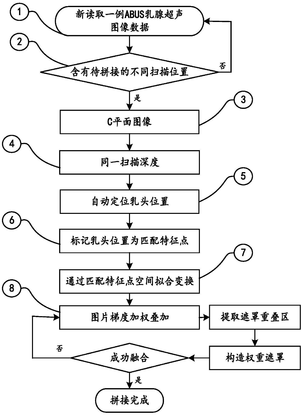
[0061] See attached figure 1 , this embodiment discloses an automatic splicing method for ABUS breast ultrasound panoramas, which can effectively and automatically stitch ABUS breast ultrasound panoramas without manual intervention, and solve the problem that the entire breast region cannot be fully presented due to the limited imaging field of view of ABUS breast ultrasound images Insufficient, doctors can general panorama Figure 1 It has a very important clinical application value to visually view the entire breast tissue structure of the examinee at one time, so as to achieve a more accurate and objective diagnosis of breast cancer screening cases.

[0062] Concrete steps of the present invention include:

[0063] An automatic stitching method for ABUS breast ultrasound panoramas, which can effectively and automatically stitch ABUS breast ultrasound panoramas without manual intervention, and solves the problem that the entire breast area cannot be fully presented due to t...

Embodiment 3

[0073]The present embodiment discloses a method for automatic splicing of an ABUS breast ultrasound panorama, comprising the following steps:

[0074] Step 1: Read a new case of ABUS breast ultrasound image data.

[0075] Step 2: Determine whether the read data contains different scanning positions to be spliced, such as: AP (front and back), Lateral (side), Medial (middle), such as Figure 4 and Figure 5 shown. If it is judged that the example data contains different scanning positions to be spliced, proceed to the next step, otherwise return to step 1 (single image does not need splicing).

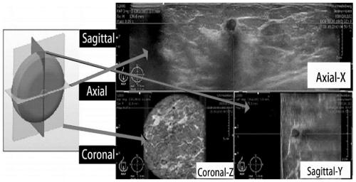
[0076] Step 3: According to the prior knowledge, automatically select the C-plane ultrasound image data from the correctly read ABUS breast ultrasound image data. An example of ABUS image data after correct reading contains image data of H plane (transverse section), S plane (sagittal plane), and C plane (coronal plane), such as image 3 As shown, in this step, the C-plane ultrasoni...


PUM


Abstract

The invention discloses an automatic splicing method and system for an ABUS mammary gland ultrasonic panorama and a storage medium. The method comprises the following steps: firstly, identifying and marking a nipple position in an ABUS image by adopting an automatic nipple position positioning method; secondly, specifying the identified and marked nipple position as a feature matching point of a subsequent splicing algorithm; fitting transformation of the to-be-spliced image is carried out through the matching feature points; and finally, superposing and fusing the pictures by using a gradientweighting method, thereby realizing automatic splicing of the ABUS mammary gland ultrasonic panorama. According to the method, the ABUS mammary gland ultrasonic panorama can be effectively and automatically spliced; manual intervention, the defect that the whole mammary gland area cannot be completely presented due to the fact that the ABUS mammary gland ultrasonic image imaging view field is limited is overcome. A doctor can visually check the whole mammary tissue structure of an examined person at a time through the panorama, breast cancer screening cases can be more accurately and objectively diagnosed, and very important clinical application value is achieved.

Description

technical field [0001] The present invention relates, in particular, to an automatic splicing method of an ABUS breast ultrasound panorama, a corresponding system and a storage medium. Background technique [0002] With the development of ABUS (automated 3-D breast ultrasound, ABUS) technology in recent years, it has gradually made up for the shortcomings of traditional two-dimensional handheld ultrasound images that are highly dependent on doctors and have a limited imaging field of view. ABUS equipment can automatically collect large volumes of information, but compared with currently used CT (Computed Tomography) and MRI (Magnetic Resonance Imaging) equipment, ABUS equipment shows a limited imaging field of view, leading to this situation The main reason is that CT and MRI can image the whole body of the patient, while ABUS imaging equipment is still limited by the size of the probe. In order to make up for this defect of the ABUS equipment, the extended field-of-view (E...

Claims


Application Information

Patent Timeline
IPC IPC(8): G06T3/40G06T7/33G06K9/32G06K9/40
CPCG06T3/4038G06T7/33G06T2200/32G06T2207/30068G06V10/25G06V10/30G06V2201/03
Inventor 吴俊耿如霞杨继婷徐丹张学杰李海燕张榆锋柏伟余韩梅
Owner YUNNAN UNIV
Why Eureka
Try Eureka