Cancer screening method based on protein marker and artificial intelligence

A protein and cancer technology, applied in the field of bioinformatics, can solve the problems of low specificity and sensitivity of tumor markers, can not meet the requirements of clinical early screening, and achieve the effect of improving accuracy

Inactive Publication Date: 2020-07-03
深圳思勤医疗科技有限公司
View PDF8 Cites 4 Cited by
  • Summary
  • Abstract
  • Description
  • Claims
  • Application Information

AI Technical Summary

Problems solved by technology

[0004] Because the specificity and sensitivity of a single tumor marker a

Method used


Image

  • Cancer screening method based on protein marker and artificial intelligence
  • Cancer screening method based on protein marker and artificial intelligence
  • Cancer screening method based on protein marker and artificial intelligence

Examples


Embodiment 1

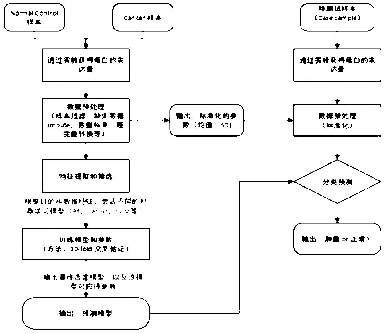
[0066] The common quantitative methods of tumor markers are: 1) Serological level uses radioimmunoassay (RIA); 2) Enzyme-linked immunoassay (ELISA); 3) Automatic immunochemical analysis system to quantify. In the present invention, the newly developed standardized method can be used to offset the impact of different quantitative methods and be used across platforms;

[0067] The samples in this embodiment are all quantified using the fully automatic immunochemical analysis system, and the specific processing process is as follows:

[0068] (1) Sample pretreatment: The blood samples of the subjects or control samples (samples of known type) are each 10ml, and the blood samples are transferred from the blood collection tube to the 10mL centrifuge tube in the biological safety cabinet, and the centrifuge tube cover is marked Sample number and date of sample processing. Centrifuge at 1600g at 4℃ for 10min. Collect the centrifuged supernatant in a new 10mL centrifuge tube. Centrifuge at...

Embodiment 2

[0072] Marker screening: using the protein tumor marker data in the published literature (Cohen, JD, etal., 2018.), the data contains protein expression data of 1817 samples, of which normal human samples: 812 cases; cancer patients Sample: 1005 cases. The data set includes: AFP, Angiopoietin-2, AXL, CA-125, CA15-3, CA19-9, CD44, CEA, CYFRA 21-1, DKK1, Endoglin, FGF2, Follistatin, NSE, OPG, OPN, PAR, Prolactin, sEGFR, sFas, sHER2 / sEGFR2 / sErbB2, etc. a total of 39 protein marker expression levels.

[0073] (1) Perform t-test comparison between cancer and normal people, and require that the amount of protein table in the cancer group is significantly higher than that in the normal population (excluding 13 protein markers, as shown in Table 1); simultaneously divide different cancers The type (only for a few cancer types with a high incidence of cancer) and the normal sample are subjected to the same hypothesis test. The more cancers with significant differences, the higher the pr...

Embodiment 3

[0086] In order to compare the prediction performance of the method developed by the present invention on early tumor screening, the inventor obtained the detection data of common tumor markers from a third-class hospital as a third-party independent verification test, which was compared with conventional clinical methods. The markers used in this example are clinically routine protein markers: AFP, CEA, CA125, CA153, CA19-9, CYFRA 21-1, etc. The validation set: includes 266 normal physical examination populations and 81 tumor patients with different cancers. Table 3 below lists the quantitative results of tumor markers in 100 samples.

[0087] table 3:

[0088]

[0089]

[0090]

[0091]

[0092] (1) Data filtering and preprocessing: For some samples in Example 1, the detection of all protein markers could not be completed for some reasons or the hospital did some special cancer-related proteins on some samples, and removed the samples. Proportion of protein marker missing> 10% ...


PUM


Abstract

The invention provides a method for determining a sample source. The method comprises the following steps: (1) carrying out protein content determination treatment on a sample so as to obtain the tumor protein marker content of the sample; (2) standardizing the sample data, wherein the data comprises the tumor protein marker content obtained in the step (1) and the dummy variable conversion data of clinical data; and (3) based on the standardized sample data obtained in the step (2) and a prediction model, determining the probability that the to-be-detected sample comes from a cancer patient.According to the method provided by an embodiment of the invention, whether the to-be-detected sample comes from a cancer patient or not can be accurately and conveniently determined through the prediction model based on the protein content and clinical data of the to-be-detected sample, so a foundation is laid for further research of cancer-related pathogenic mechanisms or early screening of cancer.

Description

Technical field [0001] The present invention relates to the field of biological information. Specifically, the present invention relates to a method for determining the source of a sample. Background technique [0002] Tumor markers are those that are synthesized, secreted and released by gene expression of tumor cells during tumorigenesis and proliferation, or are abnormally produced and / or increased by the body's response to tumor cells, which can reflect tumorigenesis, A class of substances developed to monitor tumor response to treatment. Tumor markers can be divided into: proteins; carbohydrates; lipids; enzymes; hormones, polyamines, and gene products. It exists in the tissues, body fluids and excreta of tumor patients and can be detected by immunological, biological and chemical methods. For example, radioimmunoassay (RIA) and enzyme-linked immunoassay (ELISA) and fully automated immunochemical analysis systems can be used to quantitatively detect tumor markers at the se...

Claims


Application Information

Patent Timeline
IPC IPC(8): G16B25/10G16B40/20
CPCG16B25/10G16B40/20
Inventor 李世勇茅矛张锋陈彦钟果林张岩陈灏封裕敏
Owner 深圳思勤医疗科技有限公司
Why Eureka
Try Eureka